Tiny gas bubbles, about the size of red blood cells,
can be injected intravenously, then tracked via highly sensitive ultrasound as they make their way to
tumors
Cancer cells, like healthy cells, draw their nourishment from blood — blood that is often transported
through small blood vessels that form to feed tumors. Now it appears these same blood vessels might be
harnessed to assist in diagnosing cancer, monitoring the effects of cancer treatment and even in delivering
treatment.
Tiny gas bubbles, about the size of red blood cells, can be injected intravenously, then tracked via
highly sensitive ultrasound as they travel through the bloodstream. The gas bubbles overcome a drawback
of ultrasound: While ultrasound is an excellent way of monitoring blood flow in major vessels of the body,
its use is limited in imaging very small vessels, or capillaries. That's because the relatively few red
blood cells present in capillaries don't reflect sound waves as well as surrounding tissue does. In contrast,
gas bubbles reflect a very strong, scattered signal when exposed to ultrasound, making it possible to
see their movement within almost any size of blood vessel.
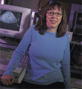
On the UC Davis campus, Katherine Ferrara,
professor and chair of biomedical engineering, has been developing sensitive methods to image bubbles
with ultrasound as they move into the capillaries of breast and prostate tumors.
The bubbles have a lipid, albumin or polymer shell and are filled either with air or certain kinds of
gases. They're attractive for use in medical research because they're unlikely to fuse, and after being
destroyed — again via ultrasound — the gases within are simply excreted through the lungs.
The techniques that Ferrara and colleagues have developed allow them to determine both bubble density
within a specific region as well as how soon a capillary will refill with bubbles once bubbles in the
vessel are destroyed with an ultrasound pulse.
"The usual speed of a capillary filling is about 1 millimeter per second," Ferrara says. "But the vasculature
in a tumor is abnormal — leaky, tortuous and not very functional, so it might take 30 seconds to refill.
The speed of refilling shows how sturdy the vessels are in that small spot. That's important because you
want to know how well chemotherapy, for example, is reaching that region of the tumor."
In addition, if gas bubbles are no longer detected in certain capillaries of the tumor following chemotherapy
or other treatment, it's a good indication that at least part of the tumor has been damaged or killed
by the therapy.
"We have confirmed that we can make very reliable and effective maps of tumors using this kind of strategy,"
Ferrara says. "We can generally pick out regions where the tumor is dead; we can even find small regions
where the tumor is still viable."
A human study of this approach may begin as early as this year in collaboration with Ralph deVere White,
director of the Cancer Center and chair
of urology; Philip Schneider, chief of surgical oncology; and John McGahan, professor of radiology.
With colleague Paul Dayton in the School of Engineering, Ferrarais also investigating another strategy:
coating the bubbles with specific molecules that cause the bubbles to adhere only to blood vessels in
tumors, not to those in healthy tissues. This is possible because blood vessels in malignant tumors possess
certain molecules not typically found in normal blood vessels . The molecules in the special bubble coating
"recognize" these tumor molecules.
Dayton has a grant from the National Institutes of Health to develop the approach, which might be used,
for example, if a mass were detected in the body and physicians needed to know whether it was cancerous.
An additional project is to make bubbles with a thicker shell — thick enough that drugs can be suspended
inside. "We're now working on locally delivering a drug to a specific region," Ferrara says. "Some of
the most toxic drugs like paclitaxel can be suspended very well."
If the bubbles are coated with a molecule that finds its match on the blood vessels of a tumor, then
the bubbles can be destroyed with ultrasound and the drug within delivered specifically to the tumor but
nowhere else.
Finally, Ferrara and her co-investigators are trying to develop a system whereby ultrasound can be used
as a force to push drug-bearing bubbles into the capillary wall of a tumor. In this scenario, the bubbles
would then be destroyed and their load released into the capillary wall — and perhaps to the tumor tissue
on the other side.
Together, the strategies have the potential to give cancer specialists new, minimally invasive and highly
targeted tools to fight cancer.