To scan or not to scan?
Researcher moves to make medical imaging safer
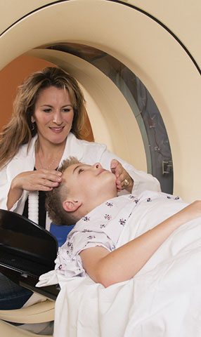
It’s a typical scenario: a young woman comes to the emergency department because of excessive vomiting and pain in her right lower abdomen.
She is examined and whisked off for a computed tomography (CT) scan, revealing an enlarged, inflamed appendix. Surgery ensues, and the patient goes home a few days later, having been cured of a potentially deadly condition.
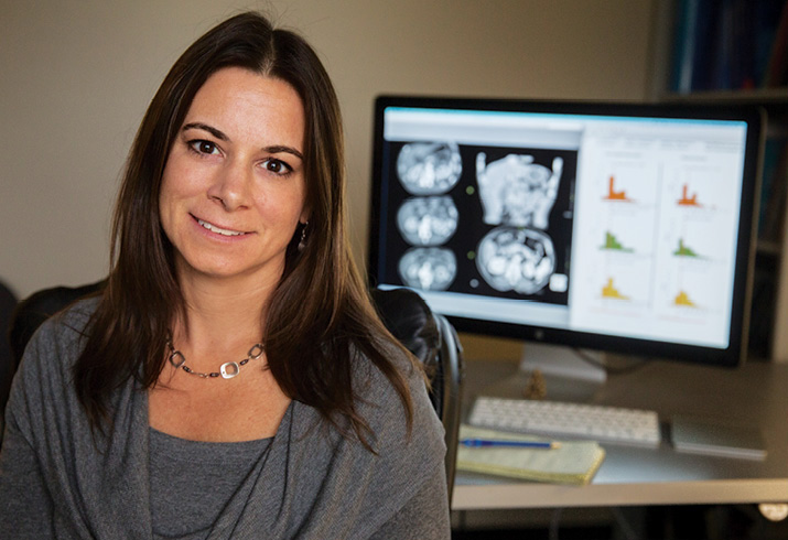
“There is no doubt that when radiographic imaging is medically necessary, the benefits outweigh the small risks from radiation exposure,” says Diana Miglioretti, Dean’s Professor in Biostatistics and a member of the UC Davis Comprehensive Cancer Center. “My efforts are directed at reducing the unnecessary exposures wherever they may occur.”
Miglioretti, who has recently been funded for three large research projects aimed at understanding and reducing radiation risk from medical imaging, is directing her sharp focus on determining whether unnecessary exposures are occurring, how dangerous they may be, and what can be done to reduce them.
Miglioretti is particularly interested in the surge in CT imaging since it became widely available a few decades ago, with an estimated 75 million scans conducted annually nationwide. The technology generates crisp three-dimensional images that allow differentiation of tissues that vary in density by less than 1 percent, enhancing the diagnosis of a host of conditions from tumors to fractures to internal bleeding. But Miglioretti cautions that the sharp images come with potential for harm: CT scanning exposes a person to up to 500 times the radiation of a conventional chest X-ray.
Focusing on pediatric cancers
About 4 million CT scans are conducted annually on children in the United States. They are speedy and convenient, and neither doctors — nor parents — want to risk missing a medical problem that should be addressed. But Miglioretti especially worries about this population of children, who, compared to adults, are most vulnerable to the long-term effects of radiation because of their rapid growth and their longer remaining lifespans.
Miglioretti and collaborators at Kaiser Permanente Northern California and UC San Francisco were recently awarded a $10.5 million, five-year grant from the National Cancer Institute to study the effects of medical radiation exposure on children. Using large databases from Group Health Cooperative, based in Seattle, Wash., and from three Kaiser Permanente regions, the research team will tally cumulative radiation exposures from medical imaging — before birth and throughout childhood — for some 7.3 million children over a 20-year period. They will search for any associated risk with the development of childhood cancers, particularly leukemia, one of the most common pediatric cancers and one that could theoretically result from radiation exposure to active bone marrow. Leukemia is easiest to study because it has the shortest lag time from radiation exposure to onset.
“We have an ideal study population and data infrastructure to do this important study,” Miglioretti says. “Because the electronic databases at each participating HMO have complete capture of all the care their enrollees receive, we can accurately estimate each child’s cumulative medical radiation exposure. This provides the best way to detect cumulative and long-term effects of tests, which on average carry a very low risk to an individual.”
She notes that while two other studies with similar aims have been published, they were based on counting the number of CT scans done on children and estimating the radiation exposure using a calculated average dose per scan. In contrast, the data that Miglioretti and her collaborators will use provide the actual radiation doses received by each individual. Miglioretti’s earlier research demonstrated that radiation doses can vary greatly across children, even for the same type of imaging test of the same anatomic region. These prior studies have been criticized because of potential confounding factors (e.g., Down syndrome, which might increase both use of CT imaging and cancer risk), and reverse causation (i.e., children may have received the CT exams because of early cancer symptoms); her study will carefully control for these potential biases.
Optimizing delivery
When Miglioretti started delving deeply into the world of medical imaging, she was shocked to discover that radiation exposure from the same type of scan can vary dramatically, depending on both the machine and its operator. No standardized protocols for CT scanning exist nationally, and although software designed to optimize dosages for different tests is available, many users opt not to buy it because of the expense.
Two additional research projects are underway to address protocols for CT scanning — a $6.2 million study funded by the National Cancer Institute, and a $1.9 million study funded by the Patient-Centered Outcomes Research Institute (PCORI). Miglioretti and her colleagues will collect detailed data to assess current practices, then develop and implement strategies to standardize and optimize CT protocols across a large number of clinics and hospitals in the United States, Canada and the United Kingdom.
“We have seen dramatic results from just discussing standardized protocols with medical physicists and radiologists in a single group meeting,” Miglioretti says. “Our studies aim to systematically find the most effective strategies, along with the most efficient ways of disseminating that information.”
Determining the optimum radiation dosages for a particular purpose can be extremely complex, she adds. The amount of radiation needed depends on the question the clinician is trying to answer; more may be needed to make an initial diagnosis than to track changes in a lesion already identified, for example. In addition, one size does not fit all; small, thin people need less radiation than large, heavy people, who require more radiation to penetrate more tissue. Children need even less.
A statistician’s perspective
Miglioretti realizes that as a statistician she looks at the problem of radiation exposure from a somewhat different angle than does an emergency department doctor or radiologist. When faced with a patient with a potential medical problem, it is natural to turn to the best test available to obtain a diagnosis if the risk of the test to the patient is low. But the risk, she emphasizes, should not be ignored, given the very large numbers of exams performed.
She wants to convince clinicians that CT imaging should not be used “just to be safe” if it is unlikely to reveal information that would change how a patient’s symptoms are managed. Ultrasound imaging, which does not use radiation, should be considered as an alternative in some situations, at least as an initial test. Ultrasound for conditions such as abdominal pain, for example, often provides results definitive enough to diagnose the problem and eliminate the need for the higher-exposure CT scan.
“Guidelines and policies for medical tests such as CT imaging consider average exposures and associated risks, but as a statistician, I worry about variability across patients,” she says. “Who are the people at the high end of the distribution getting the highest exposures? What effects might radiation have on them? What can we do to bring those high exposures down?”
With three large research grants now underway, Miglioretti is looking forward to digging into the data and finding answers.