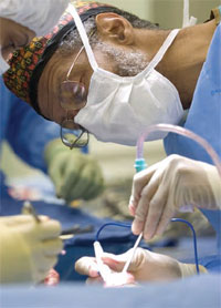
For neurosurgeons, the ability to distinguish between healthy and malignant brain tissue with pinpoint accuracy – while the patient is still on the operating table – would be a major advance. For brain cancer patients, it would mean better odds for survival.
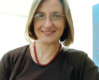
A new device under development by Laura Marcu, associate professor of biomedical engineering at UC Davis, promises to make this feat possible.
"Our method allows neurosurgeons to just shine a light on the tissue to determine whether or not it's cancer," said Marcu, who received her undergraduate degree in mechanical engineering at the Polytechnic Institute of Bucharest in Romania, her postgraduate training in spectroscopy, lasers and plasma physics at the University of Bucharest and her Ph.D. in biomedical engineering at the University of Southern California. She joined UC Davis last year from Cedars-Sinai Medical Center in Los Angeles.
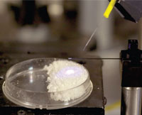
Marcu's novel technology uses laser light to excite molecules within tissues or cells. The molecules respond by fluorescing. Different molecules, depending on their chemical properties, emit light of different colors. Using sensitive optoelectronic equipment, Marcu then detects and analyzes this emitted light, gleaning important information about the biochemical status of the tissue. She also employs a process known as "lifetime" or "time-resolved" measurement to determine the wavelength of the emitted light and analyze how long it takes for the fluorescing molecules to revert to their preexcited state.
The portable set-up Marcu has created in her laboratory at the Center for Biophotonics Science and Technology at UC Davis includes a two-way fiberoptic probe that delivers an impulse of laser light and collects the re-emitted light and its decay in nanoseconds.
Precision surgery
"It is very important not to remove normal brain tissue, as this could have devastating effects on the patient. But, if tumor cells are not completely removed, the tumor will recur in a short time," Marcu said. "The techniques we have developed allow us to better identify the infiltrating tumor and the resection margin."
According to Dennis Matthews, director of the biophotonics center, Marcu's cell-screening approach promises to dramatically improve the precision of existing surgical techniques.
"Today a surgeon can take out a tumor, but can't be certain every cell of diseased tissue has been removed, hence the need for additional radiation and chemotherapy," Matthews explained. "This new technology will give surgeons the ability to survey the margins of tumors and determine with great certainty if there is any cancer left."
James Boggan, professor and vice chair of neurological surgery and co-director of the biophotonics center, said Marcu's approach has the potential to improve survival for brain cancer patients.
"Her approach is innovative and seeks to disclose heretofore hidden molecular differences between tumor cells and normal tissue," he said. "As a neurosurgeon, I am especially excited, because this work has the promise of helping me identify sub-microscopic residual brain tumor we cannot detect by our current methods. Doing so would allow us to improve the resection of brain tumors, which may help save lives."
Optical biopsy
Marcu's approach, known as time-resolved fluorescence spectroscopy and imaging, is one of several techniques under investigation at UC Davis Cancer Center for near-instantaneous optical "biopsy." A similar technology, under development by researchers at Lawrence Livermore National Laboratory in collaboration with UC Davis surgeons, is expected to enter clinical testing soon; this method relies on measurements of light scattering to distinguish normal from diseased cells.
According to Stavros Demos, a Lawrence Livermore physicist and member of the UC Davis cancer research program, optical devices for real-time tissue screening are the future of oncology.
"The current state of cancer diagnosis involves pathology, and that is a time-consuming process. It takes at least 24 hours to get the result," Demos said. "Optical biopsy technology promises to bring at least a preliminary assessment in real-time, when it would be most beneficial for both the patient and the physician."
Promising early results
Preliminary data suggest Marcu's technology will represent a significant advance in brain cancer surgery. Using her first prototype device, neurosurgeons at Cedars-Sinai Medical Center, where Marcu began her research, were able to correctly identify tumor cells left behind after the removal of glioblastomas from 17 patients. Glioblastomas are irregularly shaped brain tumors with poorly defined borders that invade neighboring tissue.
With her Cedars-Sinai collaborators, Marcu is now recruiting glioblastoma patients for a research clinical trial. The 100-patient study will also include patients being treated at UC Davis Cancer Center.
Applications in heart disease?
Marcu is also working to miniaturize her cell-screening prototype for intravascular applications, and to assess its use in cardiovascular disease. The engineer, who holds several patents, believes the approach could serve as an early warning system to detect potentially problematic atherosclerotic plaques before they can cause a heart attack or stroke.
The opportunity to take new discoveries directly to the bedside where they can benefit patients is what drew Marcu to UC Davis.
"This is a wonderful environment for bringing new technology into practice," she said. "UC Davis has the people and the resources to make new advances happen."