In translation: Ionizing radiation: a double-edged sword in cancer
 "Radiation," says Richard Valicenti, chair of the Department of Radiation Oncology at UC Davis, "is of great benefit in detecting cancer, eliminating cancer and improving survival."
But as valuable as it is in cancer diagnosis and treatment, ionizing radiation must be used carefully, because it can cause side effects such as burns and hair loss that become apparent soon after the exposure, or arise many years later in the form of secondary cancers.
Recent news reports have highlighted a few cases of radiation overdoses in clinics and added urgency to efforts to ensure the safe use of radiation in diagnosis and treatment – efforts in which clinicians and researchers at UC Davis figure prominently.
"We were looking into this problem before it got all this publicity," says Ralph deVere White, director of UC Davis' Cancer Center. "We have a lot of experts, all basically asking the same question: How can we reduce exposure while maintaining the benefit?"
Surrounded by radiation sources
Ionizing radiation can damage DNA, but our bodies are adept at performing repairs, so we can handle low exposures.
"If you can repair the cell," deVere White says, "it's not going to be mutated and you're not going to get cancer."
Low levels of radiation are naturally present in the environment: cosmic radiation from outside our solar system constantly bombards Earth; rocks and soil can contain radioactive atoms; and radiation from radon gas is a significant problem in some parts of the country.
"We get the equivalent in background radiation of a head CT every year," says John Boone, professor and vice chair of the Department of Radiology at UC Davis Medical Center, who is an expert on the physics of CT scanning and serves as chief science officer for the American Association of Physicists in Medicine.
David Rocke, a biostatistician at UC Davis, researches the effects of low-dose radiation in the environment and in medical imaging. He explains that humans have evolved mechanisms to deal with the damage that comes from exposure. He's also looking at the effects of radiation at a molecular level, using skin discarded from surgery and other procedures and an artificial skin model.
"It would be a tragedy if patients who truly needed CT scans didn't get them because they perceived that the risk was too great. "
"There are a lot of differences between individual people in their response to radiation," says Rocke. "If you know who is radiosensitive and who is not, it may influence the choice of therapies."
Ensuring patient safety
UC Davis researchers also are involved in reassessing and standardizing procedures for CT scanning and other radiation-based techniques for the hospital and clinics, as well as the industry.
At UC Davis, diagnostic and therapeutic procedures involving radiation have been reviewed to ensure patient safety, and new quality control procedures have been implemented to ensure proper functioning of machines, training of staff and thorough review of protocols for delivering radiation during diagnosis or therapy.
 "You can eliminate pretty much any cancer growth with minimal effects to the patient, but all that is very much dependent on one’s ability to apply technology to ensure the greatest degree of safety and accuracy."
Ionizing radiation was first used in medicine in 1895 when Wilhelm Conrad Roentgen produced an image of the bones of his wife’s hand, calling it an "X-ray." Later, X-rays became an invaluable tool to examine the skeletal system and to investigate problems in soft tissue, such as pneumonia.
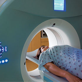
Computed axial tomography, a diagnostic imaging tool that uses X-rays, was introduced in the 1970s. CT has enabled clinicians to detect and monitor many types of cancer, as well as other diseases, and to assess the extent of injuries.
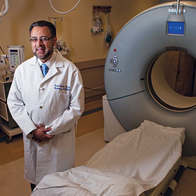
"The CT scan is one of the most important inventions of the 20th century," says Ramit Lamba, director of CT at UC Davis Medical Center and assistant professor in the Department of Radiology.
Imaging errors rare
Among imaging techniques that use ionizing radiation, CT scanning delivers the highest dose, and some researchers have suggested a link between CT scans and cancer. DeVere White, however, says many other risk factors may be involved, and notes that cancer patients, often diagnosed and monitored using CT scans, are more likely to get secondary cancers in any case.
"These studies really do raise questions, but they don't answer them," deVere White says.
As a result, Lamba says, "we have to treat CT scanning as potentially carcinogenic – without causing alarm."
Several cases of radiation overdoses from CT scans – in which human error led to incorrect doses – have made headlines recently. And while tragic, such occurrences are extremely uncommon among the 62 million CT scans performed every year, according to a 2007 study in the New England Journal of Medicine.
"CT scans are extraordinarily safe," deVere White says. "And there's absolutely no doubt that CT scans have saved a lot of lives."
 At UC Davis, diagnostic and therapeutic procedures involving radiation have been reviewed to ensure patient safety, and new quality control procedures have been implemented to ensure proper functioning of machines, training of staff and thorough review of protocols for delivering radiation during diagnosis or therapy.
Jerrold Bushberg, clinical professor of radiology and director of health physics programs at UC Davis, adds that CT scanning "has quite literally revolutionized the practice of medicine." In some cases, without it, the patient would be having exploratory surgery.
"It would be a tragedy," Bushberg added, "if patients who truly needed CT scans didn't get them because they perceived that the risk was too great."
Before ordering a CT scan, clinicians should consider if it's really necessary, says Nathan Kuppermann, chair of emergency medicine at UC Davis and co-author of an article on overuse of CT scanning in injured children, which appeared in the journal The Lancet. Although CT scans have greatly improved our ability to care for the trauma patient, CT scans should be used selectively to avoid unnecessary exposure to ionizing radiation.
If a CT scan is justified, the radiation dose should be optimized to be as low as possible while still sufficient for diagnostic purposes.
High doses required for treatment
Radiation doses delivered during diagnostic tests are small compared to those used to treat cancer, which can be thousands of times higher. Prostate cancer treatments, for example, typically involve daily doses to the prostate that total, over the course of treatment, "more than 10 times what would kill a person if their whole body was exposed to it," Rocke says.
Radiation is a valuable anti-cancer weapon because cancer cells can't repair radiation damage as readily as normal cells, and so are more susceptible.
"You can eliminate pretty much any cancer growth with minimal effects to the patient," Valicenti says, "but all that is very much dependent on one's ability to apply technology to ensure the greatest degree of safety and accuracy."
Radiation treatment used to involve a more scattershot approach, he says, but now, the radiation beam can be more accurately targeted at a tumor, so "treatments have become very precise and much more intensified," Valicenti says.
Although much of the process of radiation therapy is automated, humans still play an important role, Valicenti says. New procedures have been put in place to ensure treatment plans are peer-reviewed, and that each treatment is evaluated as if it's the first – to avoid the possibility of an error being perpetuated.
"We've reengineered and redesigned our quality assurance systems," Valicenti says, "with a focus on continuous quality control."


 John Boone: Boone is one of a handful of physicists in the U.S. specializing in computed tomography (CT) and developed one of the first dedicated breast CT scanners. He is chief science officer for the American Association of Physicists in Medicine and a co-author, with Tony Seibert and Jerrold Bushburg, of the textbook, The Essential Physics of Medical Imaging.
 Tony Seibert: Seibert is a medical physicist and president-elect of the American Association of Physicists in Medicine. He has served as an expert witness in radiation overdose cases, and is well-versed in state laws that govern radiation use. He is a member of the Alliance for Radiation Safety, which operates the Image Gently campaign, focused on reducing childhood exposure to ionizing radiation.
 James Purdy: Purdy is professor and vice chair of the Department of Radiation Oncology and director of the Physics Division. Purdy has worked with the International Commission on Radiation Units and Measurement to develop standards for specification of dose and volumes in radiation oncology.
 Sandra Gorges: Gorges is UC Davis' chief of pediatric imaging. Gorges is an expert in the radiographic evaluation of child abuse, in pediatric oncology imaging and in pediatric neuro-imaging. One of her research interests in pediatric imaging is dose reduction and appropriate utilization of resources in pediatric CT.
 Ramit Lamba: Lamba is assistant professor and chief of CT at UC Davis Medical Center. He specializes in the use of CT for diagnosis, interventional procedures and management of diseases in the abdomen and pelvis. His research focus is on the optimization of radiation dose for abdominal and pelvic CT exams.
 Nate Kuppermann: Kuppermann is professor and chair of emergency medicine and professor of pediatrics at UC Davis Children’s Hospital. He has been studying the appropriate use of CT imaging in children for more than a decade. He also is lead author of a widely-cited article recently published in Lancet on overuse of CT in children in the emergency room.
 James Holmes: Holmes is a professor in the Department of Emergency of Medicine. He conducts research on trauma patients, including studies to determine indications of abdominal CT and head CT. He can discuss balancing risks of missing important injuries versus risks of death from CT radiation-induced malignancies in both injured children and adults.
 David Rocke: Rocke is Distinguished Professor of Biostatistics in the Department of Public Health Sciences. His research explores the molecular mechanisms of the response of cells and tissues to low-dose radiation from environmental and occupational exposures. He also researches possible genetic and environmental differences among people in response to radiation.
 Jian-Jian Li: Li is a professor of radiation oncology and a researcher who studies the molecular mechanisms causing tumor resistance to radiation and chemotherapy. The goal of his work is to find therapeutic targets to enhance the cure rate for patients undergoing radiation therapy. Li also researches ways to protect normal tissue from the effects of both high-dose and low-dose radiation.
 Matthew Coleman: Coleman is associate adjunct professor of radiation oncology. He studies how low-dose radiation exposure affects gene regulation and controls cellular response. His work aims to provide the basis for identifying those factors that make cells susceptible to low-dose radiation.
 Andrew Vaughan: Vaughan is professor of radiation biology and associate chief of staff for research and development at the VA Northern California. His research centers on the fusion of certain genes that leads to development of leukemia in infants and in patients who have been exposed to radiation or chemotherapy for other cancer treatment.
 Richard Valicenti: Valicenti is professor and chair of the Department of Radiation Oncology at UC Davis. Valicenti is recognized nationally and internationally for his use of image-guidance and intensity-modulated radiation therapy in the treatment of cancer patients.
 Wolf-Dietrich Heyer: Heyer is professor of microbiology and of molecular and cellular biology and leader of the UC Davis Cancer Center’s Molecular Oncology Program. His research focuses on DNA repair, the process by which cells repair DNA and may become resistant to radiation therapy and other anti-cancer treatments.
 Stephen Kowalczykowski: Kowalczykowski is Distinguished Professor of Microbiology and of molecular and cellular biology and an elected member of the National Academy of Sciences. He is a pioneer in the analysis and visualization of DNA repair processes at the single-molecule level.


