

First-of-its-kind total-body PET scanner arrives at UC Davis
EXPLORER, the world’s first medical imaging scanner that can capture a 3-D picture of the whole human body at once, will be operational this summer. The EXPLORER Imaging Center occupies leased space on Folsom Boulevard.
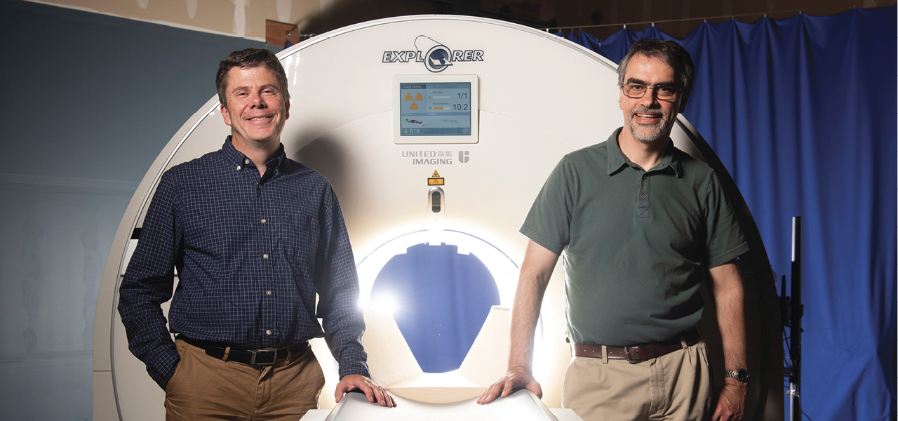
The brainchild of UC Davis scientists Simon Cherry and Ramsey Badawi, EXPLORER is a combined positron emission tomography (PET) and X-ray computed tomography (CT) scanner. Because the machine captures radiation far more efficiently than other scanners, EXPLORER can produce an image in as little as one second and, over time, produce movies that can track specially tagged drugs as they move around the entire body.
The developers expect the technology will have countless applications, from improving diagnostics and tracking disease progression to researching new drug therapies.
The scanner was developed in partnership with Shanghai-based United Imaging Healthcare (UIH), which built the system based on its latest technology platform and will eventually manufacture the devices for the broader healthcare market.
The first human scans from EXPLORER were made public in November 2018. They quickly circulated widely online, stunning radiologists and other imaging experts worldwide. The FDA approved the technology in December.
Badawi and Cherry first conceptualized a total-body scanner 13 years ago. Their idea was kick-started in 2011 with a $1.5 million grant from the National Cancer Institute and boosted in 2015 with a $15.5 million grant from the NIH.
Cherry, distinguished professor in the UC Davis Department of Biomedical Engineering, says he expects EXPLORER will have a profound impact on clinical research and patient care because it produces higher-quality diagnostic PET scans than have ever been possible. EXPLORER also scans up to 40 times faster than current PET scans and can produce a diagnostic scan of the whole body in as little as 20 to 30 seconds.
Alternatively, EXPLORER can scan with a radiation dose up to 40 times lower than a current PET scan, opening new avenues of research and making it feasible to conduct many repeated studies in an individual, or dramatically reduce the dose in pediatric studies, where cumulative radiation dose is particularly important.
For the first time, an imaging scanner will be able to evaluate what is happening in all the organs and tissues of the body simultaneously. For example, it could quantitatively measure blood flow or how the body takes up glucose everywhere in the body. Researchers envision using the scanner to study cancer that has spread beyond a single tumor site, inflammation, infection, immunological or metabolic disorders and many other diseases.
“I don’t think it will be long before we see a number of EXPLORER systems around the world,” Cherry says. “But that depends on demonstrating the benefits of the system, both clinically and for research. Now, our focus turns to planning the studies that will demonstrate how EXPLORER will benefit our patients and contribute to our knowledge of the whole human body in health and disease.”