

A new way to see cancer
Technique spares patients unnecessary surgery
Pancreatic ductal adenocarcinoma (PDAC) may be the deadliest cancer.
The five-year survival rate for patients diagnosed at stage III is 3 percent; those with stage IV disease have a 1 percent chance.
Because it is so challenging, physicians take an aggressive approach. Surgery is often indicated to eliminate the disease before it metastasizes. But these procedures move forward with limited information. In some cases, the cancer has already spread to other organs, making surgery useless. Other times, patients don’t have cancer at all.
A marker-based imaging test could profoundly impact the ability to detect pancreatic cancer before it metastasizes. In particular, this technique could benefit patients with familial pancreatic cancer or cystic lesions.
“People with familial pancreatic cancer are at higher risk, but there’s no way to tell unless you biopsy them,” says Julie Sutcliffe, professor in the UC Davis departments of Internal Medicine and Biomedical Engineering. “But you’re not going to randomly biopsy people who have not been diagnosed. Also, some patients come in with cystic lesions. These lesions may or may not become cancer, but right now we can’t tell the difference. We have to treat them like cancer, and that can lead to extra surgeries.”
No one knows this better than Richard Bold, chief of surgical oncology, who has conducted his share of PDAC procedures.
“The surgery can have significant risks,” says Bold. “Unfortunately, we convert some people to diabetics, and some of these patients might never have needed this operation. The biology of what they have is something that never would have become cancer, never would have impacted their life.”
What’s been missing is a noninvasive technique to definitively identify pancreatic tumors and gauge their spread. UC Davis researchers recently received a $3.3 million grant from the National Cancer Institute to help solve this problem. The funding will support efforts to develop advanced imaging to pinpoint pancreatic and possibly other cancers, giving clinicians more information to help patients.
Partnering to improve PET
Sutcliffe is the principal investigator on this project, but her participation highlights how circumstance can play a major role in science.
“My entry into the PDAC world was not scientific, it was personal,” says Sutcliffe. “I was diagnosed 12 years ago with breast cancer. Richard Bold was my surgeon; now he’s my collaborator.”
Along with their UC Davis colleagues, they are developing better ways to use positron emission tomography (PET) to image pancreatic tumors. PET has been a workhorse in oncology, although less successfully in pancreatic cancer. Patients receive an injection — glucose molecules attached to the radioactive isotope, fluorine 18. The idea is that cancer cells are hungrier than normal cells and will eat more glucose/fluorine molecules and light up on the scan.
That’s the theory. Unfortunately, the reality is more complicated. Normal cells and inflamed areas also can consume more glucose, and some cancers lack the protein transporters that take in blood sugar, keeping them dark in PET images.
“In a cancer patient, they may have simultaneous things going on, and we’ll have a hard time sorting out which cells are cancerous and which are not,” says Bold. “We get false positives and false negatives that really leave us in a quandary. If we could develop something that’s more specific to cancer, we could eliminate those false readings.”
Building a better target
The key to improved imaging is finding ways to get cancer cells — and only cancer cells — to take up the radioactive fluorine. This way, clinicians can be confident that the bright images on PET scans are tumors. Having this kind of specific information could be a tremendous benefit for patients.
“If the cancer has metastasized, the tumor cannot be surgically removed,” Sutcliffe says. “Unfortunately, current imaging is frequently inaccurate, and patients undergo needless exploratory surgery to identify the metastatic disease. If we could image this, we could see if the patient has disease in the pancreas, liver, lungs — wherever it has spread. We could determine if surgery will actually help the patient.”
For more than a decade Sutcliffe’s lab has been studying a molecule called αvβ6. This protein receptor lives on the invasive parts of tumors — the areas that are infiltrating healthy tissue. While αvβ6 may not be driving metastasis, it’s certainly along for the ride.
“The receptor we are targeting is expressed on pancreatic cancer cells,” says Sutcliffe. “It’s a marker for cancer aggression and not just in pancreatic cancer. There are applications in ovarian, head and neck, breast, colon, cervical and other cancers.”
Because it’s a cell surface receptor, αvβ6 offers relatively easy access to cancer cells. To take advantage, the team synthesized a peptide (a piece of a protein) that binds to the receptor. Eventually the peptide, and whatever is attached to it, is brought inside the cancer cell.
The end result is a combo molecule called 18F-αvβ6-BP, which carries a radioactive fluorine payload and binds exclusively to αvβ6. It wasn’t easy. Working with radioactive molecules requires extra care and ratchets up the difficulty level.
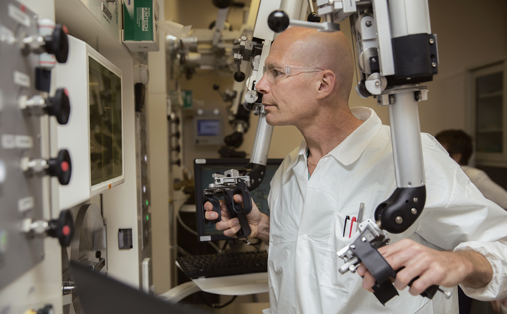
“Fluorine has a two-hour half-life, so you have to work fast,” says Sutcliffe. “It’s manufactured in the lab particle accelerator, a cyclotron, and you have to make it the day you’re using it. You can’t touch it, plus the clock is ticking.”
The process also has to meet good manufacturing practice (GMP) guidelines to ensure the quality of each batch. Still, even with these challenges, 18F-αvβ6-BP could solve a lot of problems, allowing clinicians to better assess a patient’s needs without surgery.
Joining the consortium
Sutcliffe and colleagues are excited by the prospect of helping patients with pancreatic cancer, but that’s just a start. Because αvβ6 is so common in many cancers, their new imaging molecule could be used widely to assess tumor progression.
In addition, the grant does more than fund their work; it gives UC Davis researchers access to the Pancreatic Cancer Detection Consortium (PCDC), which includes Mayo Clinic, Johns Hopkins University, Dana Farber Cancer Institute and other institutions.
“We become the major imaging site and can recruit patients from the other sites,” said Sutcliffe. “This is a really dynamic group of investigators, and we believe we can make a lot of progress toward an effective early-detection method.”