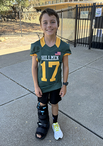
When playing football, Ryder Phippin got hit hard, like so many times before. He pushed through the pain and finished the game, but his leg pain continued intermittently. His parents took him to see a doctor a couple months later.

His doctor figured Ryder’s injury was most likely a calcified contusion, a condition where bone tissue forms within a muscle after a bruise. He recommended rest, ice and physical therapy, which is usually effective in managing symptoms.
“He told us it would just go away eventually,” said Jennifer Phippin, Ryder’s mother. But the little bulge and hard spot in the skin kept getting worse.
By this time, Ryder was in excruciating pain, especially at night. On a return visit to the doctor, the medical team took an X-ray and the results were alarming.
“It was a surreal moment,” Jennifer said, recalling how she heard the doctor say, “osteoid osteoma.” “I didn’t know what that meant. She just kept saying ‘it’s not cancer, it’s not cancer.’ I was very confused by that and I was thinking, ‘You mean it could have been?’”
An osteoid osteoma is a benign bone tumor that is often characterized by injury to the area where the tumor occurs. The main symptom is pain which often gets worse at night. It was exactly what Ryder had experienced.
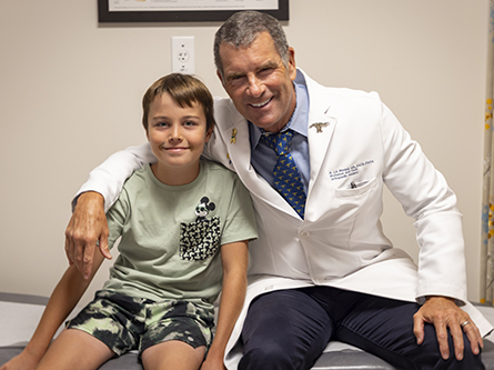
Ryder needed a pediatric orthopaedist. He was connected to UC Davis Health and to R. Lor Randall, a renowned pediatric surgeon and oncologist and also the chair of the Department of Orthopaedic Surgery at UC Davis Health.
“Osteoid osteomas aren’t typically that large, but his tumor was very large, over a centimeter and growing,” Jennifer recalled the doctor explaining. “He also said the tumor wasn’t showing up on the X-ray like a normal osteoid osteoma does.”
Despite the challenges, Randall assured the Phippins he could likely perform a CT-guided radiofrequency ablation, a modern procedure that burns out the tumor.


