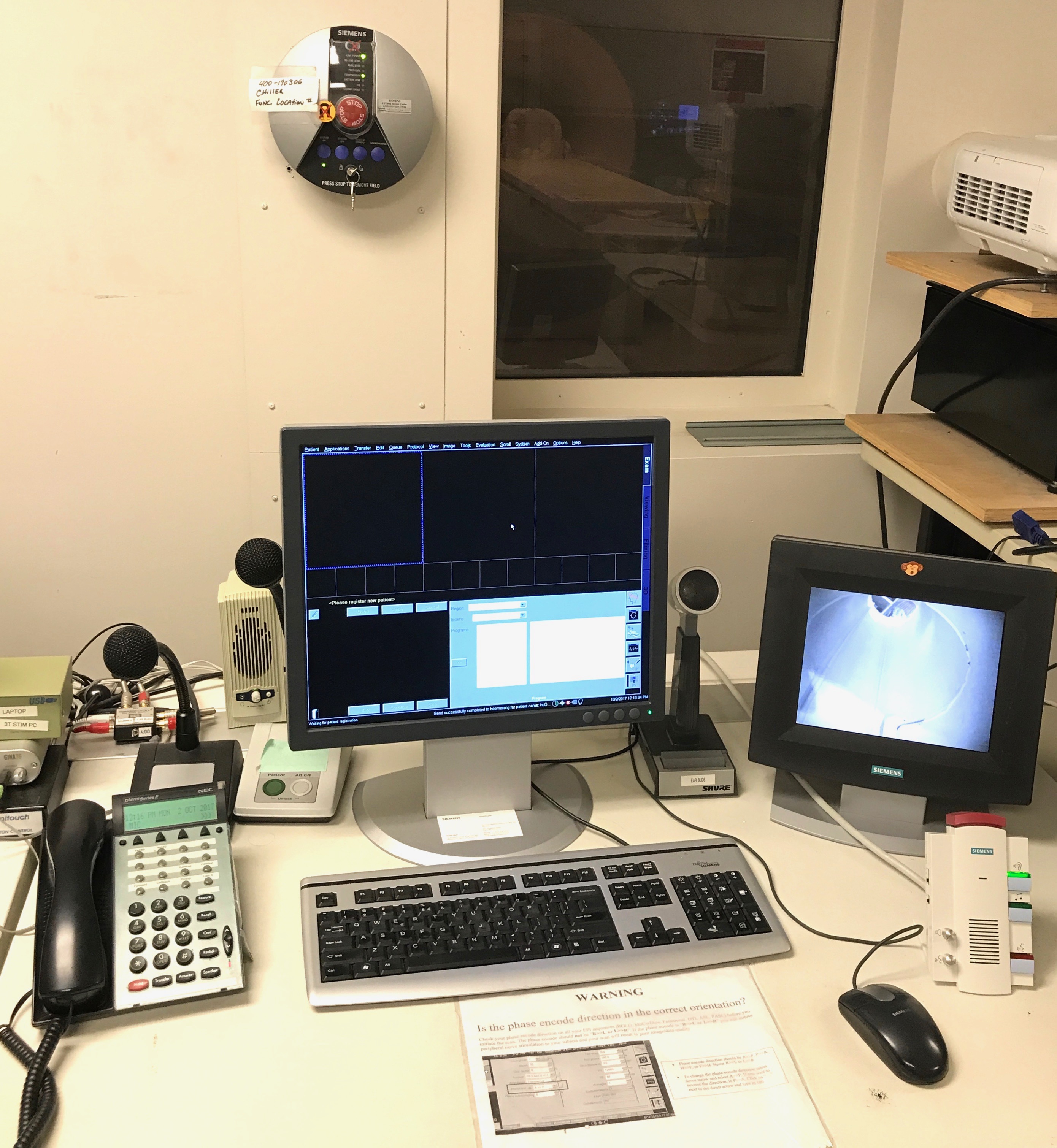
Peripheral Equipment

Visual and Auditory Presentation Systems
Visual System
The visual stimulus presentation systems for the Siemens Prisma 3T scanner have been designed to present high resolution in-bore images with the tightly controlled timing necessary for recording event-related potentials (ERP) simultaneously with fMRI data acquisition. We achieve this with the BOLDscreen 24 LCD for fMRI, by Cambridge Research Systems.
The BOLDscreen incorporates a high resolution 1920 x 1200 IPS LCD panel with native 8-bit color resolution, providing a true-color 16.7 million color display and contrasts of up to 1000:1. The BOLDscreen is fully MRI compatible and is mounted at the rear of the bore, where the 24" LCD display provides the maximum field of view when sited at the rear of the bore and viewed via headcoil mounted mirrors.
Auditory System
Auditory stimuli are presented during scanning via two high-fidelity systems designed for the MR environment: Optoacoustics OptoACTIVE noise-canceling headphones and Sensimetric model S14 insert earphones.
The Optoacoustics headphones provide active and passive EPI gradient noise reduction to provide clear sound for audio stimulus. They also have a slim profile for use with 32 and 64 channel head coils. These headphones are CE MDR certified and US FDA 510(k) cleared.
The S14 Insert Earphones are used when high quality sound is needed in a restricted space. They use piezo-electric transducers and Comply foam canal tips to achieve both a flat frequency response ( from 100Hz – 8 kHz ) and high attenuation of scanner noise.
Eye-Movement Monitoring Equipment
For the 3T MRI system, the eye-tracking system is the EyeLink 1000 Plus long-range, manufactured by SR Research of Ottawa, Canada. The system includes a dedicated computer running a real time operating system, a fiber optic camera, long range infrared illuminator and an MRI safe mounting system. The eye-tracker system is monocular and can record data at a rate of 1000 Hz. The data recorded includes the X, Y position, saccade onsets and offset times, and pupil size with an accuracy of .25-.50 degrees and a resolution of .05 RMS and .25 degrees saccade resolution. At 1000 Hz, the real-time data access has a mean of 2.2 ms with a SD of 0.3 ms. The eye-link system has broad software support. The IRC has used the eye-tracker in projects using the stimulus presentation software E-Prime, Matlab, and Presentation.
Stimulus Presentation and Button Response Recording Equipment
On the 3T system, investigators use Windows workstations to deliver visual paradigms. Stimulus presentation and button-press response acquisition is controlled by the e-Prime (Psychology Software Tools, Inc.) or Presentation software system (Neurobehavioral Systems, Inc.), Psychophysics Toolbox (open source toolbox for Mathworks), or PsychoPy (open source Python). Subjects respond with button presses obtained with specially constructed MRI-compatible fiber-optic devices (fORP by Current Designs). There are several response devices available, 8 buttons (4 each hand), 5 button (single hand) and joy stick. The metal-free response devices are connected via fiber optic cable to an optoelectronic controller unit. Note! All the above equipment can also be used with lab provided laptops with the OS and software of their choice.
Physiological Monitoring and System Diagnostics Equipment
Physiological waveforms from the subject being scanned can be recorded using the BioPac MP 150 system. Available modules include cardiac, respiratory, galvanic skin response, and other physiological signals from the subject. The Siemens Tim Prisma offers a built-in physiological measurement unit (PMU). Using MRI safe Bluetooth transmitters, the PMU system can capture pulse-ox and respiration data with a resolution of 20 ms and 3 lead EKG data with a resolution of 2.5 ms.
Mock MRI System
The simulated environment of an MRI system, consisting of a mock-up of an MRI system, non-functioning RF coils, and including a built-in speaker system for playing back recordings of the pulse sequence sounds. This environment also includes the visual and auditory stimulus presentation systems that are used in the actual MRI environment. Using the mock MRI system, potential subjects for the MRI scanning sessions can be acclimated to and trained for fMRI studies. A headphone-mounted motion detector trains subjects to become aware of their own head motion, which must be minimized during an actual scan.
