
Research
Imaging
Mission
 Musculoskeletal disease conditions are complex and multifaceted. Medical imaging provides unique, powerful tools to interrogate musculoskeletal tissue on a structural, functional and molecular level. These tools therefore offer the ability to better understand musculoskeletal disease and generate improved treatment strategies. The mission of the Imaging Core of the Center for Musculoskeletal Health is to:
- Develop and utilize a wide range of imaging technologies for humans and small animals directed toward problems of musculoskeletal health,
- Synergize with and provide resources for musculoskeletal research within the center and at UC Davis, and
- Translate the knowledge gained into improved understanding and better treatment of musculoskeletal diseases.
Facilities and resources:
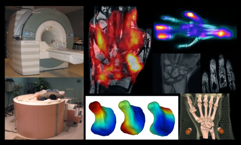
The Imaging core actively collaborates with the Center for Molecular and Genomic Imaging (CMGI), the Imaging Research Center, the Department of Radiology and the Center for Visualization at UC Davis. Investigators at the Center of Musculoskeletal Health have access to two 3T and two 1.5 T MRI scanners for use in humans and primates, a 7T small-animal MRI scanner, a wide range of X-ray and CT scanners, two PET/CT human whole-body scanners, a SPECT/CT human whole-body scanner, two microPET systems, a microSPECT/CT system, two advanced optical imaging systems, and a range of ultrasound systems through this collaboration. In addition, new instrumentation being developed by the Imaging core members for high resolution extremity imaging is also available to the team. A wide range of test objects (phantoms) for calibration, characterization and quality control of imaging system are available. The Imaging core has access to a dedicated 60-CPU core PC cluster with approximately 2000 GPU cores and 30 Terabytes of RAID storage for computationally intensive work. A range of image processing software is also available, including ITK/VTK, MATLAB, MedINRIA, 3D Slicer, Rview and ImageJ.
Imaging research conducted as part of the Center for Musculoskeletal Health
Selected current projects:
- Development of dedicated high-resolution in vivo imaging systems capable of PET/CT or Optical/Ultrasound imaging of the human and animal musculoskeletal system
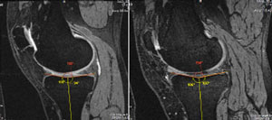
- Protocol development for performing MRI of the extremities and the carpal tunnel during continuous active joint motion
- Morphometric analyses of bones and soft tissue of the musculoskeletal system
- Development of advanced image processing methods for performing spatial alignment of bones and soft tissue using quasi-rigidity constraints.
- Monitoring of response to biologics in human rheumatoid and psoriatic arthritic patients using extremity PET/CT and SPECT/CT
For more information, please contact Abhijit J. Chaudhari, Ph.D. or Nancy Lane, M.D.
