
MR Physics
Audrey Fan
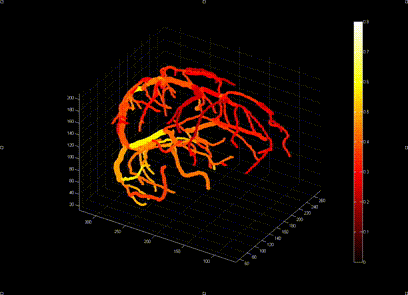
Dr. Audrey Fan develops novel imaging methods to measure cerebral physiology, including brain oxygenation, cerebral blood flow, and vascular reactivity. She has designed advanced quantitative susceptibility mapping algorithms to visualize the brain’s vasculature and its oxygenation state, based on deoxyhemoglobin concentrations in blood. Dr. Fan also has quantitative modeling experience to understand microvascular function in brain tissue from the BOLD (blood oxygenation level dependent) MRI signal.
Dr. Fan is currently engineering advanced vascular “fingerprinting” techniques to image multiple brain physiological parameters from a single, fast sequence using a dictionary matching approach. She is passionate about multi-modal imaging and how the synthesis of MRI with positron emission tomography (PET) can inform meaningful biomarkers of brain function.
Youngkyoo Jung
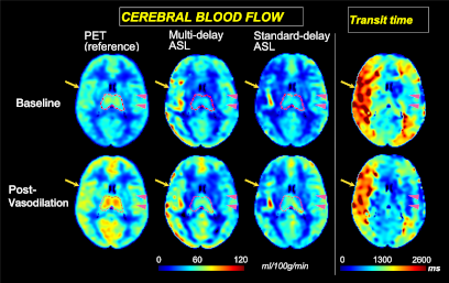
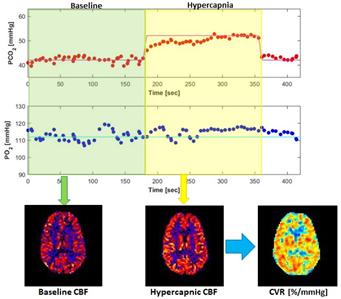
Dr. Jung's research work has focused on the development of novel structural and functional neuroimaging techniques, which have resulted in a variety of innovations, and the application of structural and functional imaging methods to study normal development/aging and a variety of diseases affecting the central nervous system. His current research includes an investigation of cerebrovascular contribution to cognitive decline. The figure below describes a technique he has developed for vascular reactivity measurement using a hypercapnia paradigm, which represents vascular function. Blood flow maps are obtained during baseline and hypercapic periods and per-voxel based cerebrovascular reactivity images are generated.
Abhijit Chaudhari
Abhijit J. Chaudhari, Ph.D., is an Associate Professor of Radiology and Director of the Center for Molecular and Genomic Imaging at University of California Davis, and a core scientist at the California National Primate Research Center. Four primary areas of research his lab pursues are:
- real-time MRI acquisition and reconstruction schemes;
- high-performance, low-field MRI optimization;
- MRI-based image segmentation, registration and morphometry; and
- quantitative multimodality biomarkers.
His current projects are related to imaging of the musculoskeletal and neurological system. His lab receives grant support from the National Institutes of Health, the National Science Foundation and the National Psoriasis Foundation. Lab website: https://chaudharilab.faculty.ucdavis.edu/
Felipe Godinez
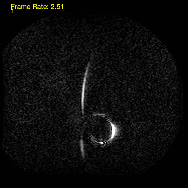 Dr. Godinez’s research focuses on the development of novel medical imaging techniques, with emphasis on treating disease in the brain and the heart. The development of these techniques is enabled through fundamentally advancing Magnetic Resonance (MR) and Positron Emission Tomography (PET) imaging instrumentation, acquisition, and image and data processing. His current projects include the use of parallel transmit MR for the safe visualization of endovascular devices and the development of MR imaging protocols for real-time interventional procedures in the heart and brain. He is interested in developing interventional MR methods that support targeted drug delivery in neuro-oncologic treatments. He is also co-developing a PET insert for simultaneous PET/MR imaging for small animals. He is interested in the combined use of PET/MR to explore MR based therapeutic biomarkers in neuro-oncologic disease.