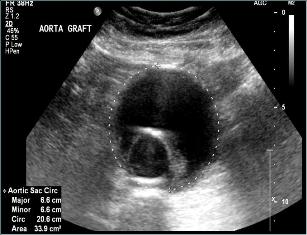
Abdominal aortic aneurysm endograft

Duplex imaging of a treated aneurysm with endograft device
While treatment of abdominal aortic aneurysm (AAA) with an endograft — also known as an endoluminal graft or stent-graft — is a much less invasive approach to treating AAA, endografts have specific technical limitations and potential complications that must be considered. Once an endograft has been placed, there is a need for long-term surveillance to ensure that the graft remains in the correct position, that flow through the graft is normal and that the aneurysm is not expanding.
An "endoleak" is present if there is flow in the aneurysm sac outside the endograft. Some endoleaks require treatment, others can be observed.
The schedule for post-operative surveillance imaging may vary, but usually tests are ordered at three, six and 12 months after the endograft is placed, then annually thereafter. Plain X-rays are used to look at the stent positioning and a CT scan with contrast is used to evaluate for the size of the aneurysm and endoleak. A duplex scan in the Vascular Laboratory will be ordered at the first follow-up visit; and if ultrasound imaging provides a complete assessment, duplex scans will be substituted for CT scans at the time of subsequent evaluations.
Ultrasound duplex scanning is non-invasive and does not require use of X-ray contrast. The duplex scan can show the size of the aneurysm sac, the absence or presence of endoleak, and it can assess blood flow through the endograft.
Some preparation is needed. The study examines arteries in the abdomen and pelvis. Gas in the intestinal tract can interfere with ultrasound evaluation. It is therefore best to have the examination performed after an overnight fast and it is important to avoid tobacco and caffeine prior to the test. In addition to a complete duplex scan of the implanted graft, the aorta and the iliac arteries, ankle and arm blood pressures will be measured using inflatable cuffs and an ultrasound Doppler flow detector. A complete study can take two hours.
