


Fomina Lab
Physiological Effects of T Cell Exosomes
Although activated T cell secrete large amounts of exosomes into the extracellular space, their physiological role remains unclear. Because T cell exosomes contain raft-associated GM1 glycosphingolipids and expose phosphatidylserine (PS) on their outer membrane leaflet they may be targets for professional phagocytes, such as monocytes and macrophages, which express PS receptors and participate in inflammatory response.
Recruitment of a large number of monocytes and lymphocytes to the arterial wall is a hallmark of atherosclerosis. During the development of an atherosclerotic lesion, macrophages accumulate massive amounts of cholesterol esters and are converted to foam cells. Apoptosis of foam cells is a major feature in the formation of the lipid-rich necrotic core associated with late atherosclerotic plaques. In advanced human plaques, T cells constitute approximately 10% to 20% of the cell population, and they are often located at the site of atheroma rupture. The role of T cells in development and progression of atherosclerosis remains controversial. The purpose of this study was to examine whether T cell exosomes could affect lipid accumulation in human macrophages.
We found that exposure to T cell exosomes enhances production of proinflammatory cytokine TNF-α and promotes monocyte lipid accumulation. (See Figure 6 below.)
Because monocytes and activated T cells are abundant within the atherosclerotic plaque, our results indicate that exosomes released by atheroma-associated T lymphocytes may accelerate atherogenesis due to stimulation of monocyte lipid accumulation and pro-inflammatory response. Due to the apparent significant role of the T cell exosomes in the development and progression of cardiovascular diseases and stroke, we currently focus our efforts on identifying the molecular mechanisms regulating exosome biogenesis and secretion.
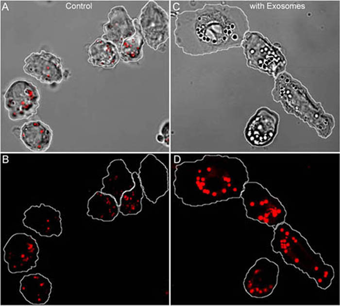
Figure 6. T cell exosomes stimulate lipid accumulation in human monocytes.
(A-D), confocal images of human monocytes after overnight incubation in the absence (A,B) or presence (C,D) of T cell exosomes. Red staining corresponds to Nile Red fluorescence specific to neutral lipids. In A and C, Z projections of the fluorescent stacks are overlaid on transmitted light images. Note that brightest Nile Red fluorescence originates from the spherical droplets.
Because monocytes and activated T cells are abundant within the atherosclerotic plaque, our results indicate that exosomes released by atheroma-associated T lymphocytes may accelerate atherogenesis due to stimulation of monocyte lipid accumulation and pro-inflammatory response. Due to the apparent significant role of the T cell exosomes in the development and progression of cardiovascular diseases and stroke, we currently focus our efforts on identifying the molecular mechanisms regulating exosome biogenesis and secretion.
 Make a donation using our secure online system.