• Simó Lab

    Research Programs

  • Simó Lab

    Research Programs

  • La Torre Lab

    Research Programs

  • La Torre Lab

    Research Programs

Hana Anderson, Ph.D.

Hana Anderson, Ph.D.

Associate Professor
hnanderson@ucdavis.edu

Dr. Anderson's research is on medical education. She is interested in developing and disseminating undergraduate medical education materials in foundational sciences such as genetics and gross anatomy as well as educational theories and pedagogies.

Nadean Brown, Ph.D.

Nadean Brown, Ph.D.

Professor
nlbrown@ucdavis.edu

The Brown lab uses mouse and in vitro models to investigate the genetic pathways underlying mammalian eye formation. This research will contribute to a better understanding of congenital eye diseases and ultimately inform stem cell therapies to correct vision loss. Currently, our primary research focus is to understand the earliest steps of ocular morphogenesis, optic nerve head formation, and astrocyte specification and differentiation.

Marie Burns, Ph.D.

Marie Burns, Ph.D.

Professor
meburns@ucdavis.edu

The Burns lab examines the biochemical and biophysical properties of signaling in photoreceptors, as well as the consequences of defective signaling on visual performance and degeneration. Our work also examines the gliosis and neuroinflammation that results from photoreceptor degeneration with the goal of mitigating photoreceptor stress and cell death.

Fernando A. Fierro, Ph.D.

Fernando A. Fierro, Ph.D.

Associate Adjunct Professor
ffierro@ucdavis.edu

The Fierro lab primary works on altering gene and microRNA expression levels of human MSC to either optimize their therapeutic potential for applications such as bone repair, non-healing ulcers and critical limb ischemias or to understand the basic mechanisms involved in differentiation, proliferation, self-renewal, etc.
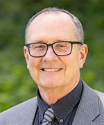

Paul FitzGerald, Ph.D.

Paul FitzGerald, Ph.D.

Distinguished Professor and Chair
pgfitzgerald@ucdavis.edu

The FitzGerald lab has identified two proteins that are very divergent members of the intermediate filament family of proteins that assemble into a unique cytoskeletal element called the beaded filament. Both of these proteins and the beaded filament are expressed only in the lens.
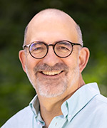

Tom Glaser, MD Ph.D.

Tom Glaser, M.D., Ph.D.

Professor
tmglaser@ucdavis.edu

The Glaser lab studies genetic mechanisms of vertebrate eye development, evolutionary conservation of gene networks, and transcriptional regulation. The lab's major interests include early patterning of the optic primordia and retinal cell fate specification.
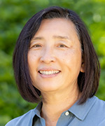

Qizhi Gong, Ph.D.

Qizhi Gong, Ph.D.

Professor
qzgong@ucdavis.edu

The Gong lab is interested in the molecular mechanisms regulating olfactory system development as well as pathogenesis in neurodegenerative disease models. We use mouse models and combine with current molecular genetics, genomics, and imaging approaches to investigate innate immune capacity and neuroinflammatory responses in the olfactory system.
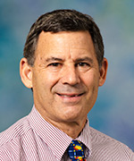

Douglas S. Gross, M.D., Ph.D.

Douglas S. Gross, M.D., Ph.D.

Senior Lecturer
dsgross@ucdavis.edu

Dr. Gross's research includes integrated management of childhood illness, tropical medicine and international health
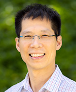

Henry Ho, Ph.D.

Henry Ho, Ph.D.

Associate Professor
hyhho@ucdavis.edu

The Ho lab has focused on an evolutionarily conserved and clinically relevant signaling pathway called the Wnt5a-Ror pathway. The Wnt5a-Ror pathway regulates tissue morphogenesis as well as other developmental and regenerative processes, and dysfunction of the pathway causes a broad range of human diseases, including structural birth defects, neurodevelopmental disorders and metastatic diseases.


Li-En Jao, Ph.D.

Li-En Jao, Ph.D.

Associate Professor
ljao@ucdavis.edu

The Jao lab is interested in understanding how the centrosome and its related organelles such as the cilium coordinate signals that regulate cell function and influence development.


Paul Knoepfler, Ph.D.

Paul Knoepfler, Ph.D.

Professor
knoepfler@ucdavis.edu

The Knoepfler lab is interested in epigenetics in stem cells and cancer. We use cutting edge molecular, cellular and developmental biology methods as well as genomic and gene editing technologies to answer key open questions in these areas of research. We are particularly interested now in the roles of three factors normally in stem cells and during tumorigenesis: histone variant H3.3, the MYC family, and DPPA4. How do these factors link the epigenome to cellular behaviors and tissue growth?
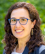

Anna La Torre, Ph.D.

Anna La Torre, Ph.D.

Associate Professor
3311 Tupper Hall,
Davis Campus
530-752-9103
alatorre@ucdavis.edu

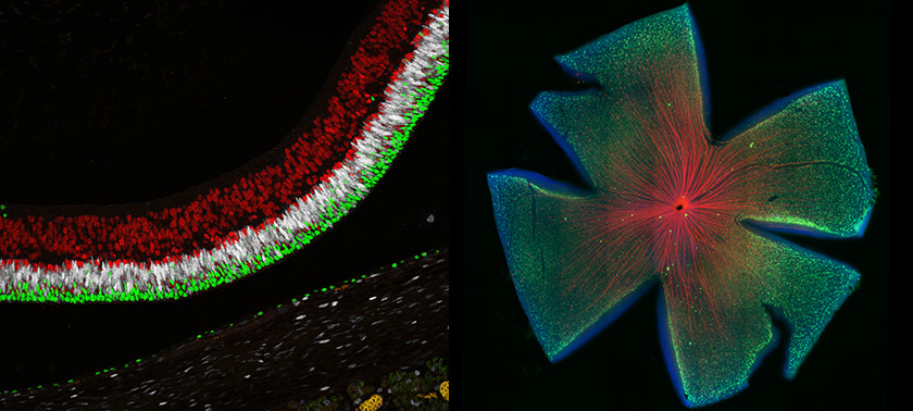
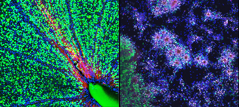
The La Torre lab studies different aspects of retinal development, including retinal patterning, neurogenesis, and foveal development to devise novel treatments to restore vision. We use different animal models and stem cell-derived organoids in combination with molecular and histological approaches and imaging techniques.
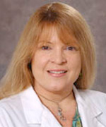

Jan Nolta, Ph.D.

Jan Nolta, Ph.D.

Professor
janolta@ucdavis.edu

The basic research in the Nolta laboratory focuses on understanding the dynamics of stem cell migration and attraction to sites of injury. Following intravenous infusion, adult stem cells home to sites of tissue damage. Areas studied are cellular response to hypoxia and chemokines, cell motility, cell-to-cell interactions, and paracrine factors secreted by MSC at the site of injury.
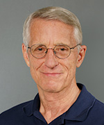

Edward N. Pugh, Jr., Ph.D.

Edward N. Pugh, Jr., Ph.D.

Distinguished Professor
enpugh@ucdavis.edu

The Pugh lab studies phototransduction, the molecular and biophysical mechanisms by which rod and cone photoreceptors in the retina transduce light into electrical signals. In the past decade it has focused on phototransduction in mouse cone photoreceptors.
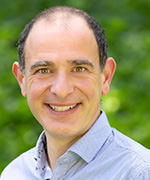

Sergi Simó, Ph.D.

Sergi Simó, Ph.D.

Associate Professor
ssimo@ucdavis.edu

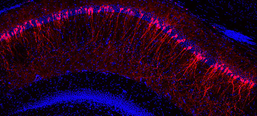
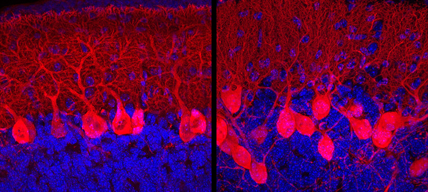
The Simó lab studies the understanding of normal brain development to address developmental and neurodegenerative disorders


Alice F. Tarantal, Ph.D.

Alice F. Tarantal, Ph.D.

Professor
aftarantal@ucdavis.edu

The Tarantal lab’s research program includes the following areas of translational research:

  • Gene therapy and somatic cell gene editing
  • Stem and progenitor cell therapies / Regenerative Medicine
  • Fetal: Pediatric models of human disease
  • Fetal: maternal microchimerism and precision models
  • In vivo translational imaging applications
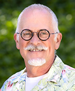
Richard P. Tucker, Ph.D.

Richard P. Tucker, Ph.D.

Distinguished Professor
rptucker@ucdavis.edu

The Tucker lab studies cell-cell and cell-extracellular matrix interactions, especially how they relate to cell motility and differentiation. He also studies the evolution of extracellular matrix and cell-cell adhesion molecules. Model systems used include the development of the avian visual system, tumor metastasis, stem cell niches and growth and regeneration in the marine invertebrate Nematostella. Most of his work focuses on the roles of tenascins, thrombospondins and teneurins. Tenascins and thrombospondins are extracellular matrix glycoproteins that can influence cell motility, proliferation and differentiation in the embryo. They often reappear in tumors and at sites of trauma. Teneurins are transmembrane glycoproteins expressed by interconnected populations of neurons. Recent evidence suggests that teneurins evolved via horizontal gene transfer between a choanoflagellate and prokaryotic prey.


Sailabala Venkata Vanguri, M.D.

Sailabala Venkata Vanguri, M.D.

Assistant Adjunct Professor
svvanguri@ucdavis.edu

Dr. Vanguri is experienced in teaching Gross Anatomy and its allied fields like Histology, Neuroanatomy, Embryology, and Radiological Anatomy.

Megan Y. Dennis, Ph.D., (D.Phil.)

Megan Y. Dennis, Ph.D., (D.Phil.)

Assistant Professor
Department of Biochemistry and Molecular Medicine
mydennis@ucdavis.edu

Dr. Dennis's research focuses on identifying genes and variants contributing to neurodevelopment impacting unique human cognitive features as well as disorders, including autism and epilepsy. Her group employs innovative genome sequencing approaches to identify variants/genes as well as functional genomics and model organisms (namely, zebrafish) to understand their functions in neurodevelopment.


Brian Leonard, D.V.M., Ph.D.

Brian Leonard, D.V.M., Ph.D.

Assistant Professor
Surgical and Radiological Sciences
School of Veterinary Medicine
bcleonard@ucdavis.edu

As a comparative ophthalmologist, Dr. Leonard's research is focused on the physiology and pathobiology of the ocular surface, including infectious keratitis, corneal fibrosis, corneal biomechanics and innate immunity, as well as spontaneous and experimental models of ophthalmic disease.


Nicholas R. Marsh-Armstrong, Ph.D.

Nicholas R. Marsh-Armstrong, Ph.D.

Professor
Department of Ophthalmology and Vision Science
nmarsharmstrong@ucdavis.edu

Dr. Marsh-Armstrong's laboratory studies cellular and molecular mechanisms of relevance to glaucoma. The laboratory's main interests are in the roles of mitochondria dysfunction within retinal ganglion cells, the role of astrocytes in clearing debris from the optic nerve head, and the potentially deleterious inflammatory response at the optic nerve head.


Ala Moshiri, M.D., Ph.D.

Ala Moshiri, M.D., Ph.D.

Professor
Department of Ophthalmology and Vision Science
amoshiri@ucdavis.edu

The Moshiri's lab seeks to advance novel treatments to the clinic for his patients. The majority of his research work centers on retinal cell replacement through transplantation of photoreceptors to the retina and gene therapy approaches for photoreceptor diseases. He also studies retinal embryology and genetics to understand the molecular underpinnings of macular development, survival, and function.


Paul A. Sieving, M.D., Ph.D.

Paul A. Sieving, M.D., Ph.D.

Professor
Department of Ophthalmology
pasieving@ucdavis.edu

Dr. Sieving’s research focuses on working with patients who have genetic retinal diseases and on developing laboratory approaches to repairing injured retinal cells and tissues. He works to understand eye diseases toward developing therapies for inherited retinal dystrophies. He established the Center for Ocular Regenerative Therapy (CORT) at UC Davis Health in 2020. CORT physicians and scientists are developing gene and stem cell therapies for these conditions. He currently is conducting a human gene therapy clinical trial for X-Linked Retinoschisis (XLRS) disease. Dr. Sieving holds elected membership in the National Academy of Medicine USA, in 2006, and in the German Academy of Science in 2013. He has published more than 250 scientific papers and book chapters. He previously was Director of the National Eye Institute at NIH.


Sara M Thomasy, D.V.M., Ph.D.

Sara M Thomasy, D.V.M., Ph.D.

Professor
Surgical and Radiological Sciences
smthomasy@ucdavis.edu

The Thomasy lab focuses on teh corneal wound healing, large animal models of ophthalmic disease, ocular pharmacology and toxicology, glaucoma, and antiviral therapy for feline herpesvirus


Glenn C. Yiu, M.D., Ph.D.

Glenn C. Yiu, M.D., Ph.D.

Professor
Department of Ophthalmology
gyiu@ucdavis.edu

The Yiu lab employs a combination of retinal biology and ocular imaging technologies across various animal models including mice, monkeys, and human patients. By developing optically-activated drug delivery systems, optogenetic disease models, and gene editing strategies, their lab hopes to better understand the pathophysiology of retinal disorders, and discover novel therapies for their patients.


Robert J. Zawadzki, Ph.D.

Robert J. Zawadzki, Ph.D.

Associate Professor
Department of Ophthalmology and Vision Science
rjzawadzki@ucdavis.edu

Dr. Zawadzki has a broad background in biomedical optics, biomedical engineering, and vision science. He is currently developing several novel in vivo retinal imaging modalities to study structural and functional changes of the retina over time at cellular resolution. These include several multimodal (combined OCT and fluorescence SLO) adaptive optics enhanced clinical and basic science retinal imaging systems. Additionally, he has experience in the development and application of early generation of OCT handheld based retinal imaging systems for clinical investigations in the pediatric population. He is also involved in development of novel image acquisition and data processing schemes that help to quantify structural and functional changes in the retina in response to normal aging, disease progression as well as therapy. As an example, one of the new functional retinal tests developed at UC Davis, so-called optoretinography (ORG), focuses on measuring light-evoked optophysiology signals from photoreceptors using OCT. We choose term ORG to show similarities to ERG (electroretinogram), which has long been used to assess retinal function in vivo.