Fetal echocardiograms
Download Fetal Echocardiograms information sheet
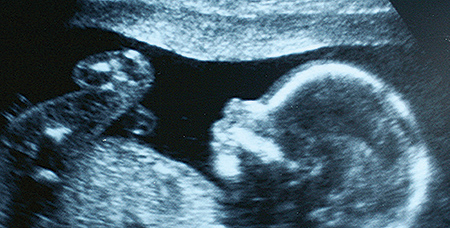
This test is used to look at the structure, function and rhythm of the heart. The ultrasound is performed by a specially trained fetal cardiac ultrasound sonographer and the images are immediately interpreted by a doctor with specialty training in pediatric cardiology and fetal congenital heart disease.
Certain women are at a higher risk of having a baby with congenital heart disease. Current recommendations for a fetal echocardiogram include:
- In vitro fertilization (IVF) or assisted reproductive technology (as published, up to 2x the risk)
- Concerns on a routine obstetrical ultrasound
- Increased nuchal translucency
- Family history of congenital heart disease (1st degree relative)
- Abnormality of another organ system
- Abnormal fetal heart rate or rhythm
- Known or suspected fetal genetic condition
- Monochorionic twin pregnancies
- Abnormal maternal blood screening
- Insulin dependent diabetes mellitus
- Maternal phenylketonuria (PKU)
- Maternal autoimmune disease (lupus)
- Maternal use of certain medications
- Exposure to certain viral infections
- Presence of hydrops
When should I schedule a fetal echocardiogram?
The best gestational age for a fetal ultrasound is between 18-22 weeks. However, fetal echocardiograms can be performed as early as 16 weeks and throughout your pregnancy.
What are the limitations of fetal echocardiogram?
There are some heart abnormalities that cannot be detected prenatally including mild valve abnormalities and small holes between the chambers of the heart. There are also some heart defects that are not evident until after the baby is born.
What happens after a fetal echocardiogram?
After the ultrasound, the doctor (fetal cardiologist) will immediately give you the results. If the ultrasound is normal, typically no other testing is needed. If heart defects are seen on the ultrasound additional testing will be recommended. This may include further echocardiograms, genetic testing and consultations with other doctors (fetal surgeon, pediatric cardiothoracic surgeon, perinatologist, neonatologist).


