Residency Program - Case of the Month
August 2009 - Presented by Phillip Starshak, M.D.
Clinical history:
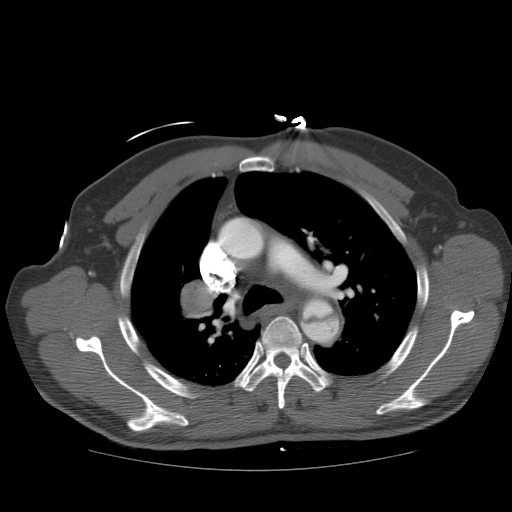
The patient is a 63 year-old male who presented to the emergency room because of sharp chest pain radiating to the scapula. A chest x-ray revealed a widened mediastinum and a CT angiogram confirmed a type B dissection of the thoracic aorta. Incidentally found was a well-circumscribed enhancing mass in the upper right hilum measuring 2.4 x 2.8 cm. A subsequent PET/CT scan was done which demonstrated hypermetabolism of this mass with a SUV of 5.7 but no areas suspicious for metastatic disease. The patient was taken to the OR for resection of this mass via right lobectomy.
CT scan image
Gross description:
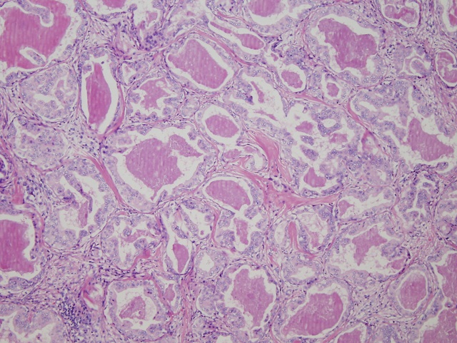
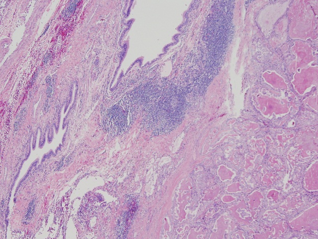
At the hilum was a well-circumscribed round shaped mass measuring 3.4 x 3.0 x 2.6 cm with tan-white cut surfaces. The mass protruded in to the lumen of the bronchus (no gross photos available).
Immunohistochemical stains:
- Thyroglobulin: Negative
- Synaptophysin Negative
- TTF-1: Negative
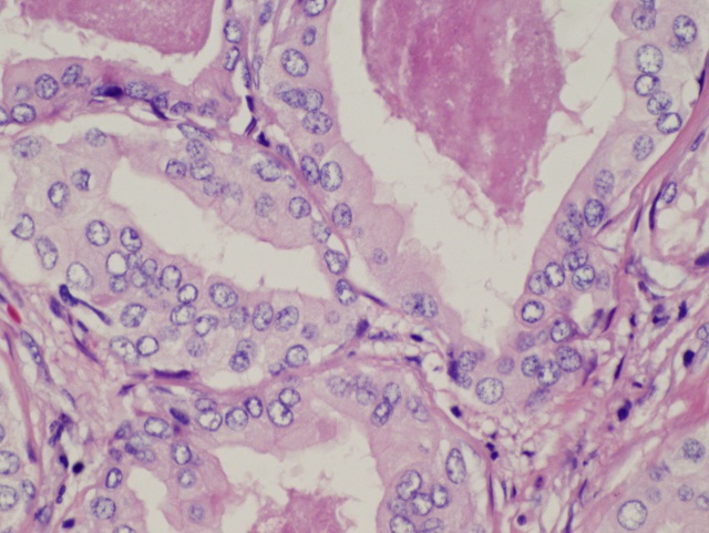
- SMA: Highlights virtually all the nests of cells
Special stains:
- PAS with diastase: Positive
- Mucicarmine: Positive


 Meet our Residency Program Director
