Residency Program - Case of the Month
April 2010 - Presented by Aram Millstein, M.D.
Clinical history:
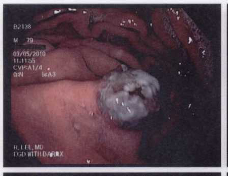
The patient is a 79 year old man with a history of Barrett's esophagus. A surveillance EGD one year prior, demonstrated an area of high grade dysplasia which was subsequently treated with radiofrequency ablation. He presented for a repeat EGD with possible radiofrequency ablation to areas of Barrett's esophagus. The EGD revealed two 0.5 cm diameter pigmented polyps in the stomach, one on the greater curvature and the other on the lesser curvature. These two polyps were completely separate and discrete. Both were snared, removed, and submitted to Pathology. They are described below.
Gross description:
Both polyps were noted to be tan polypoid soft tissue fragments, measuring roughly 0.7 x 0.6 x 0.6 cm and 0.7 x 0.7 x 0.5 cm. The polyps were bisected to reveal tan-brown and brown cut surfaces. (No gross photos available).
EGD photograph:
H and E Slides:
(The two polyps had identical histology)


 Meet our Residency Program Director
