Residency Program - Case of the Month
July 2010 Final Diagnosis - Presented by Tatyana Berezenko, M.D.
Answer:
Epithelial-myoepithelial carcinoma
Histological description
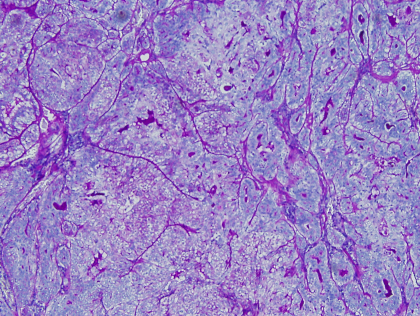
The tumor demonstrates a multinodular growth pattern and is surrounded by well-defined fibrous capsule. The salient feature of this tumor is that it demonstrates a biphasic appearance. This biphasic appearance shows a dual population of cells with one population composed of small cuboidal cells with amphophilic cytoplasm and eccentric small nuclei that form ductlike structures with central lumen formation containing acellular eosinophilic material. These cells represent the ductal epithelial cell component. The other population of cells is composed of oval, polyhedral cells with clear cytoplasm which represent myoepithelial cell component. The ductal epithelial cells contain varying amounts of glycogen in their cytoplasm that can be demonstrated by a PAS stain. In addition, the lumens that these cells form is often filled with acellular deeply eosinophilic material that stains positive for PAS-D and is thought to represent basement membrane material. The mitotic rate is low in this tumor with a few mitotic figures in the clear-cell component.
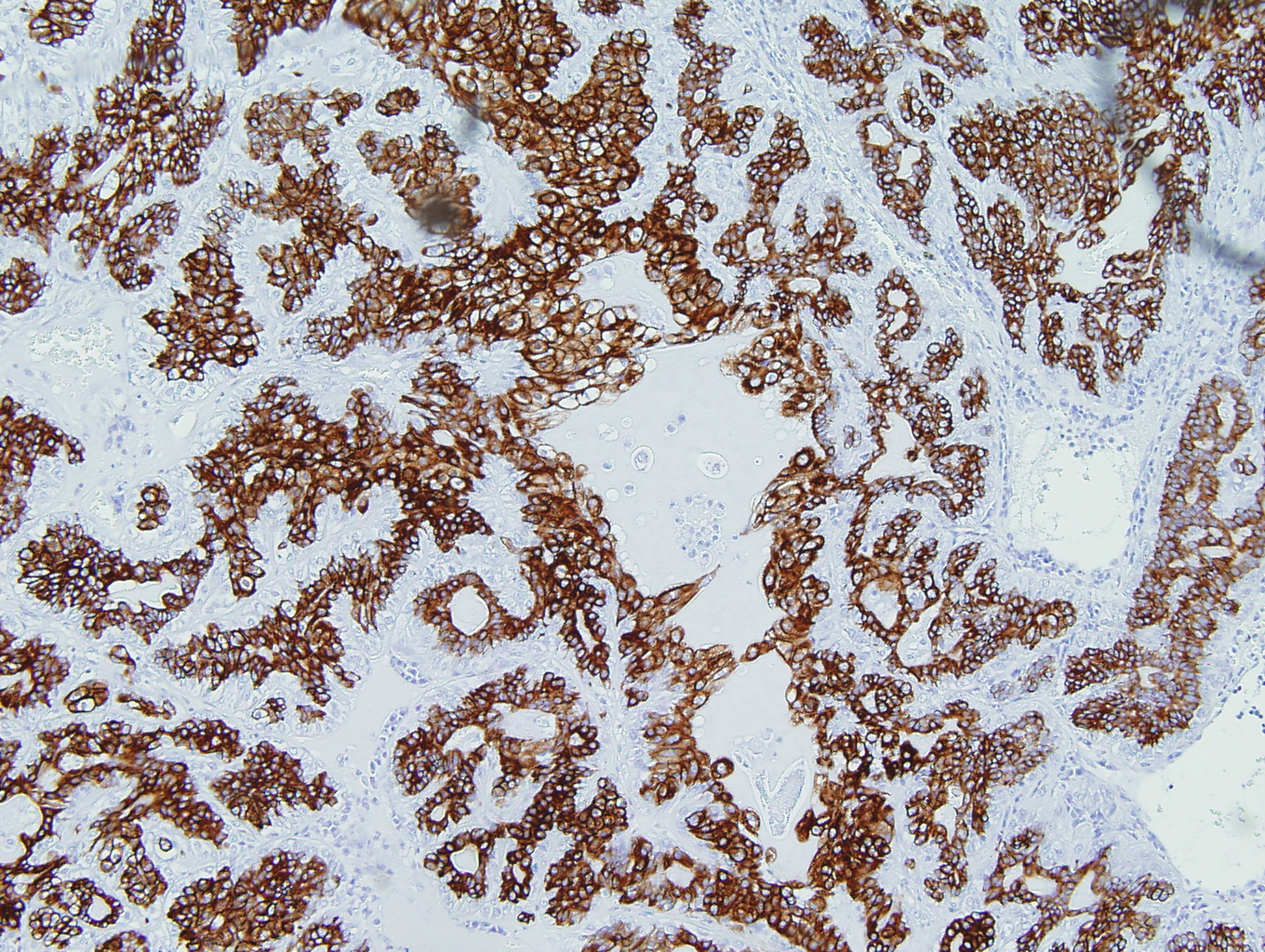
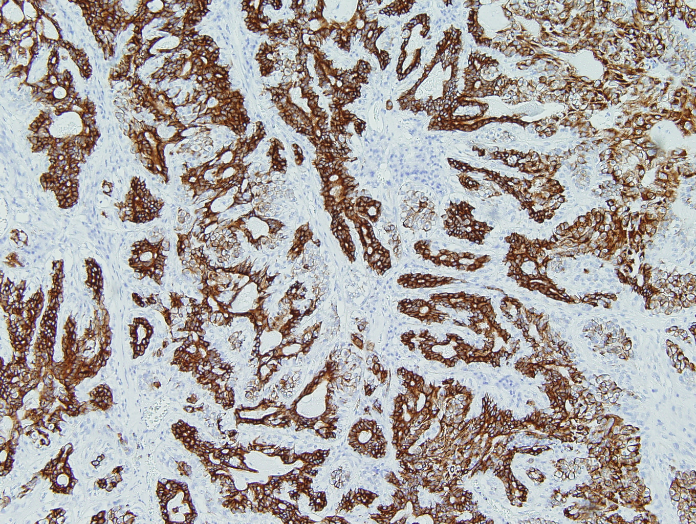
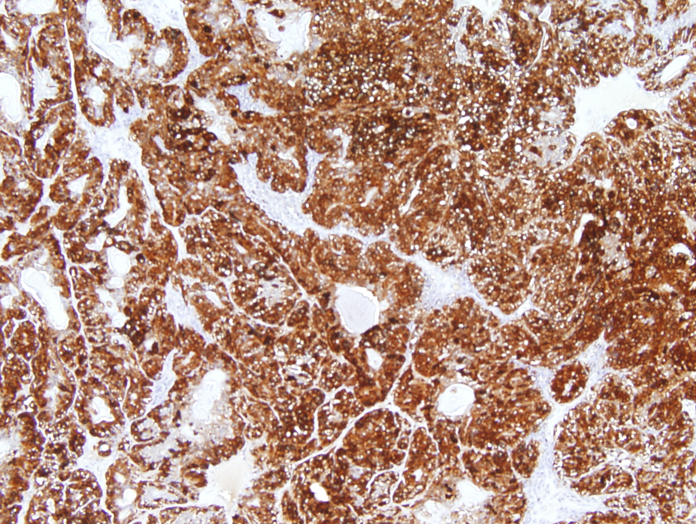
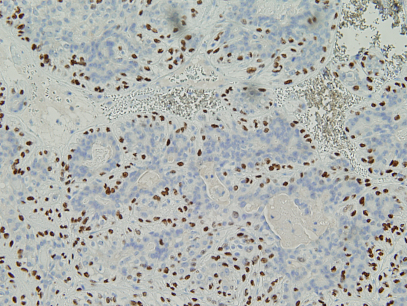
This biphasic/dual population is highlighted even more dramatically by immunohistochemical stains. The ductal epithelial cells react with cytokeratins (low-molecular weight cytokertains) and the clear cells (myoepithelial cells) demonstrate positive reactivity for high-molecular weight cytokertains (34BE12) and other myoepithelial cell markers (S-100, p63, CD10, SMA, HMA) while the ductal component is negative for these stains, with the one exception being S-100 which shows weak staining in the ductal epithelial component while the myoepithelial cells shows strong staining.
Immunohistochemistry
|
Figure 1: PAS-D - highlights intraluminal basement membrane material |
Figure 2: CAM5.2 - positive in epithelial cells |
|
Figure 3: 34BE12 - positive in myoepithelial cells |
Figure 4: S100 - positive in the myoepithelial component and weakly positive in the epithelial cell component |
|
Figure 5: P63 - highlights myoepithelial cell component |
|
Discussion:
The epithelial-myoepithelial carcinomas represents less than 0.5% of all salivary gland neoplasms and can often be underdiagnosed and even misdiagnosed in cases with a predominance of either the myoepithelial or ductal epithelial component. In such cases that show a predominance of one cell type thorough sampling of these tumors is important to demonstrate the two cell population. Tumors occur in patients usually in their mid-60s and are twice as common in women then in men. More than 80% occur in the parotid gland while the rest occur in the submandibular and minor salivary glands. Slow progressive swelling with or without facial nerve palsy are the cardinal symptoms at presentation. Epithelial-myoepithelial carcinomas behave as a low grade malignancy with a high likelihood for local recurrence. Regional lymph node metastases and distant blood-borne metastatic spread to lungs and kidneys have been reported.
Histologic sections of these tumors usually show a well-defined fibrous capsule surrounding a multinodular growth pattern. Not infrequently, the capsule is focally absent or is breached by nests of tumor cells. As mentioned earlier this tumor is characterized by two cell types producing the biphasic appearance. One population is composed of small cuboidal cells with amphophilic cytoplasm and eccentric small nuclei form ductlike structures with central lumen formation and eosinophilic material representing basement membrane that is PAS-D positive. Surrounding these cells is a layer of larger oval or polyhedral myoepithelial cells with clear cytoplasm on H&E-stained sections. Hyalinization and cyst formation may be prominent features of this tumor and sometimes is related to a prior fine needle aspiration procedure. The mitotic rate is usually low, with most mitotic figures noted in the clear-cell component. Mucin production is not evident in these tumors.
In tumors having the above sterotypical appearance, the diagnosis is straight forward. However, in some epithelial-myoepithelial carcinomas, the ductal component can be difficult to spot in H&E-stained sections and is often hidden in an overgrowth of the clear-cell, myoepithelial cell component making the diagnosis difficult. The differential diagnosis of epithelial-myoepithelial carcinomas includes several primary salivary gland tumors most notably pleomorphic adenoma, myoepithelioma and clear cell carcinoma, not otherwise specified (NOS). Pleomorphic adenomas tend to form well-defined, ovoid or round tumors. In addition, pleomorphic adenoma often show spindled shaped cells within a stromal component composed of myxoid and chondroid like substance which you do not see in epithelial-myoepithelial carcinomas. Another differential diagnosis would be myoepithelioma. Myoepithelioma is composed almost exclusively of sheets, islands or cords of cells with myoepithelial differentiation that may exhibit spindle, plasmacytoid or clear cytoplasmic features without an epithelial component. The cells of myoepithelioma are usually positive for high-molecular weight cytokeratins and myoepithelial cell markers but do not stain for low-molecular weight cytokeratins where as epithelial-myoepithelial carcinomas tend to stain positive with low-molecular weight cytokeratins and high-molecular weight cytokeratins. Differential diagnosis with clear cell carcinoma, not otherwise specified relies on the demonstration of the peculiar amyoid-like quality of the stroma with focally positive staining for low-molecular weight cytokeratins and negative for myoepithelial cell markers. Clear cell carcinomas are also unencapsulated and infiltrative.
Whenever attempting to diagnose this entity metastatic clear-cell carcinoma of the kidney should also be given serious diagnostic consideration but clinical history will often be helpful in this context.
References:
- "Epithelial-myoepithelial carcinoma: a review of the clinicopathologic spectrum and immunophenotypic characteristics in 61 tumors of the salivary glands and upper aerodigestive tract." Seethala RR, Barnes EL, Hunt JL.
- "Epithelial-myoepithelial carcinoma of the lung: immunohistochemical and ultrastructural observations and review of the literature." Wilson RW, Moran CA.
- "Myoepithelial tumors of salivary glands: a clinicopathologic, immunohistochemical, ultrastructural, and flow-cytometric study." Alós L, Cardesa A, Bombí JA, Mallofré C, Cuchi A, Traserra J.
- "Diagnostic criteria for neoplastic myoepithelial cells in pleomorphic adenomas and myoepitheliomas. Immunocytochemical detection of muscle-specific actin, cytokeratin 14, vimentin, and glial fibrillary acidic protein." Takai Y, Dardick I, Mackay A, Burford-Mason A, Mori M.
- "Epithelial-myoepithelial carcinoma: An unusual tumor of the paranasal sinus." Sunami K, Yamane H, Konishi K, Iguchi H, Takayama M, Nakai Y, Wakasa K.

 Meet our Residency Program Director
