
Residency Program - Case of the Month
February 2011 - Presented by Rebecca Sonu, M.D.
Clinical history:
The patient is a 66 year-old female with a past medical history of hypertension, atrial fibrillation and congestive heart failure who underwent a computed tomography (CT) scan of the abdomen to evaluate for an existing abdominal aneurysm. The final radiology report resulted in an incidental finding of several splenic nodules within an enlarged spleen with the largest nodule measuring up to 2.2 cm in diameter (Figure 1). The positron emission tomography (PET) scan showed unremarkable metabolic activity for malignancy, flow cytometry results did not demonstrate monoclonal activity, and fine needle aspiration showed normal red pulp of the spleen. To rule out malignancy, the patient underwent laparoscopic splenectomy for final pathologic diagnosis.
|
Figure 1. Computed tomography scan of abdomen. Enlarged spleen containing multiple hypodensities of varying size with the largest measuring 2.7 cm. |
||
 |
||
Gross description:
On gross examination, the splenectomy consisted of multiple fragments of macerated spleen weighing 190 grams and measuring 13.0 x 10.2 x 3.5 cm in aggregate. The parenchyma is beefy red and soft with no areas of consolidation (Figure 2).
|
Figure 2. Fragment of the splenectomy specimen. |
||
 |
||
Microscopic Photographs:
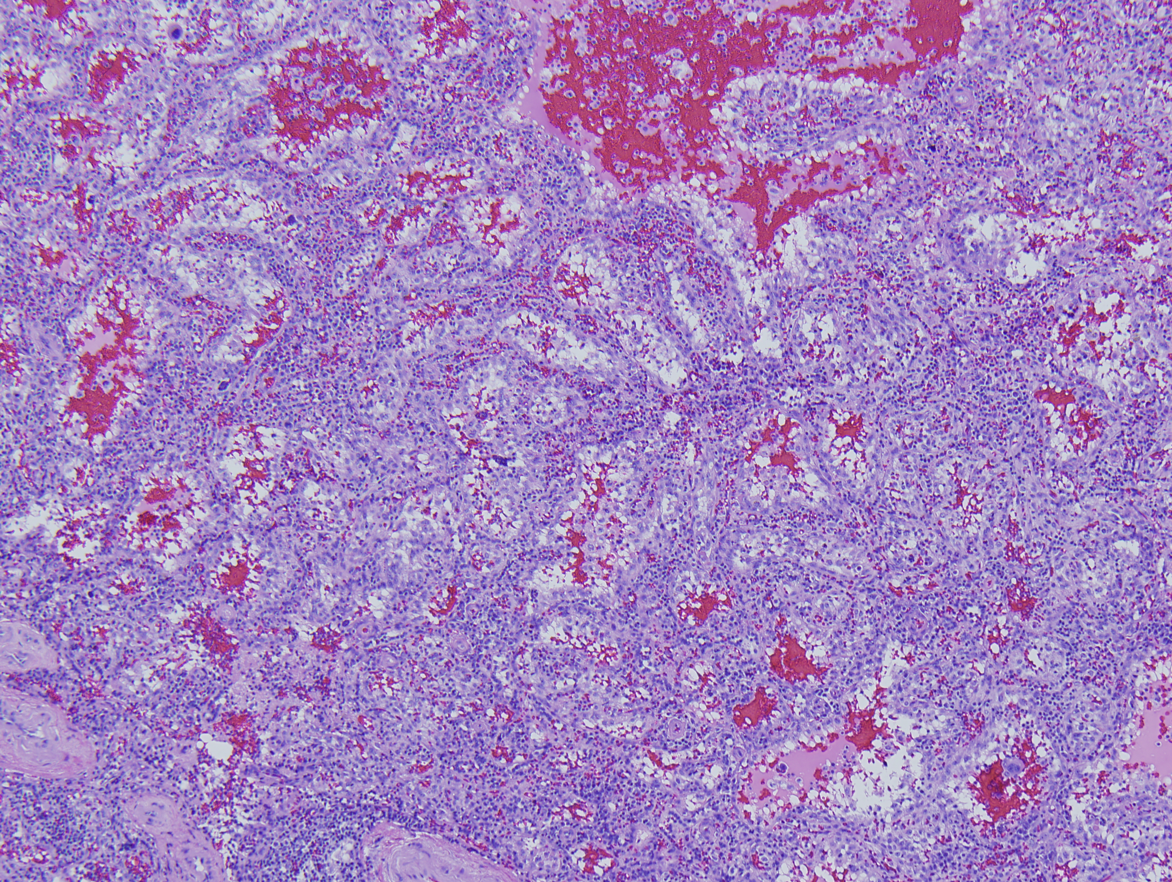 |
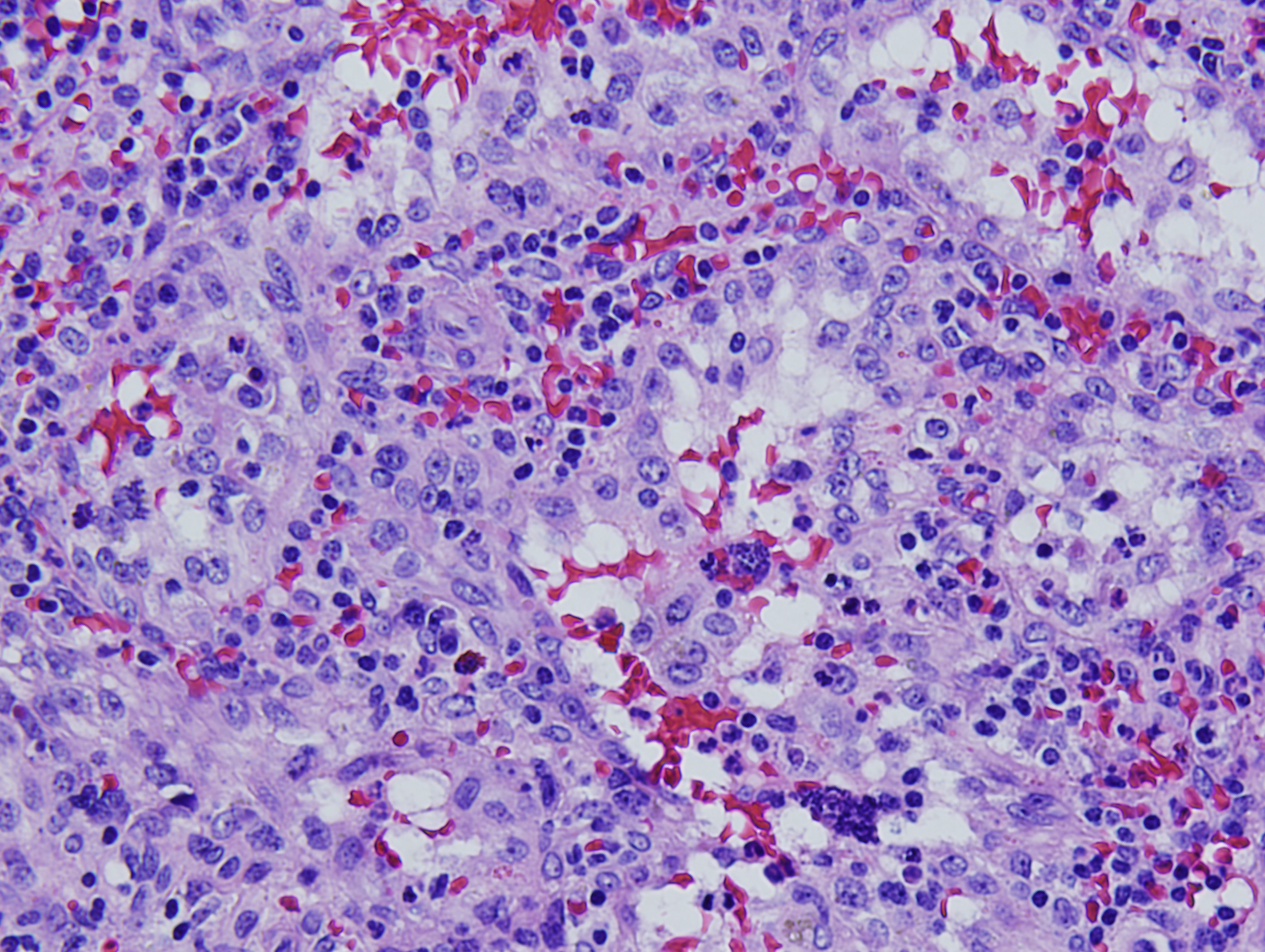 |
 Meet our Residency Program Director