
Residency Program - Case of the Month
April 2011 - Presented by Aram Millstein, M.D.
Clinical history:
The patient is a 38 year old male with a history of a left testicular varicocele and a chief complaint of infertility for four years. He had no other significant past medical or surgical history. A scrotal ultrasound done as part of the work up for infertility, showed a 0.5 cm hypoechoic mass in the right testicle, along with the varicocele in the left testicle. The radiologist noted that the right testicular mass was worrisome for a seminoma. A right radical orchiectomy was performed and the orchiectomy specimen is described below.
Gross description:
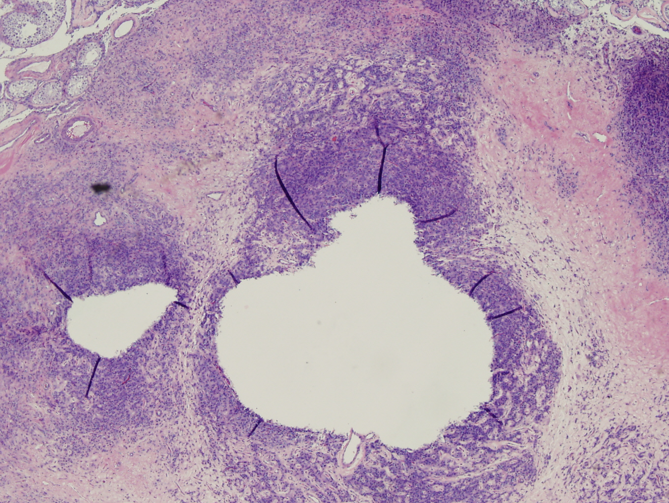
The right orchiectomy specimen consisted of a 4.2 x 2.2 x 1.9 cm testicle with attached 8 cm long spermatic cord. The tunica vaginalis was intact. Sectioning of the testicle revealed spongy, brown to yellow testicular parenchyma containing a 0.5 cm pale area. The histology of this area is shown below.
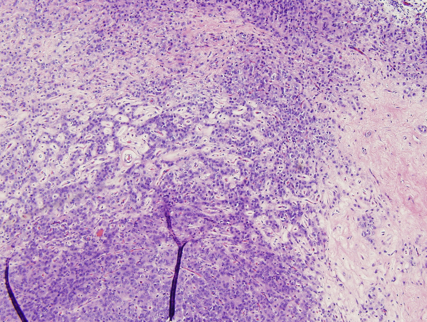
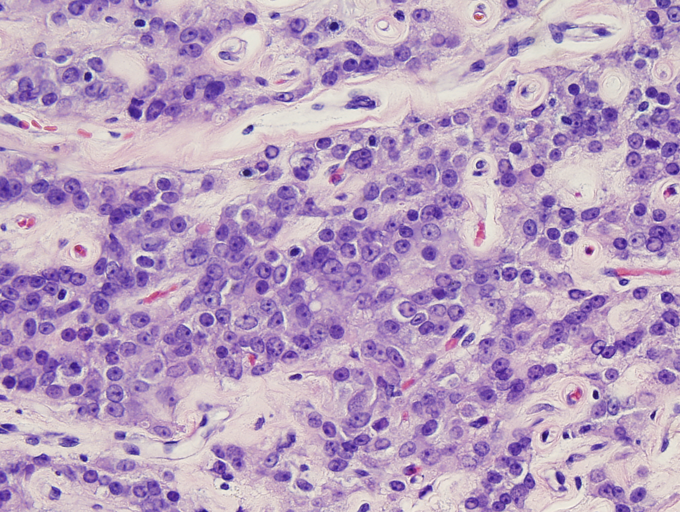
Microscopic photographs:
 |
 |
 |
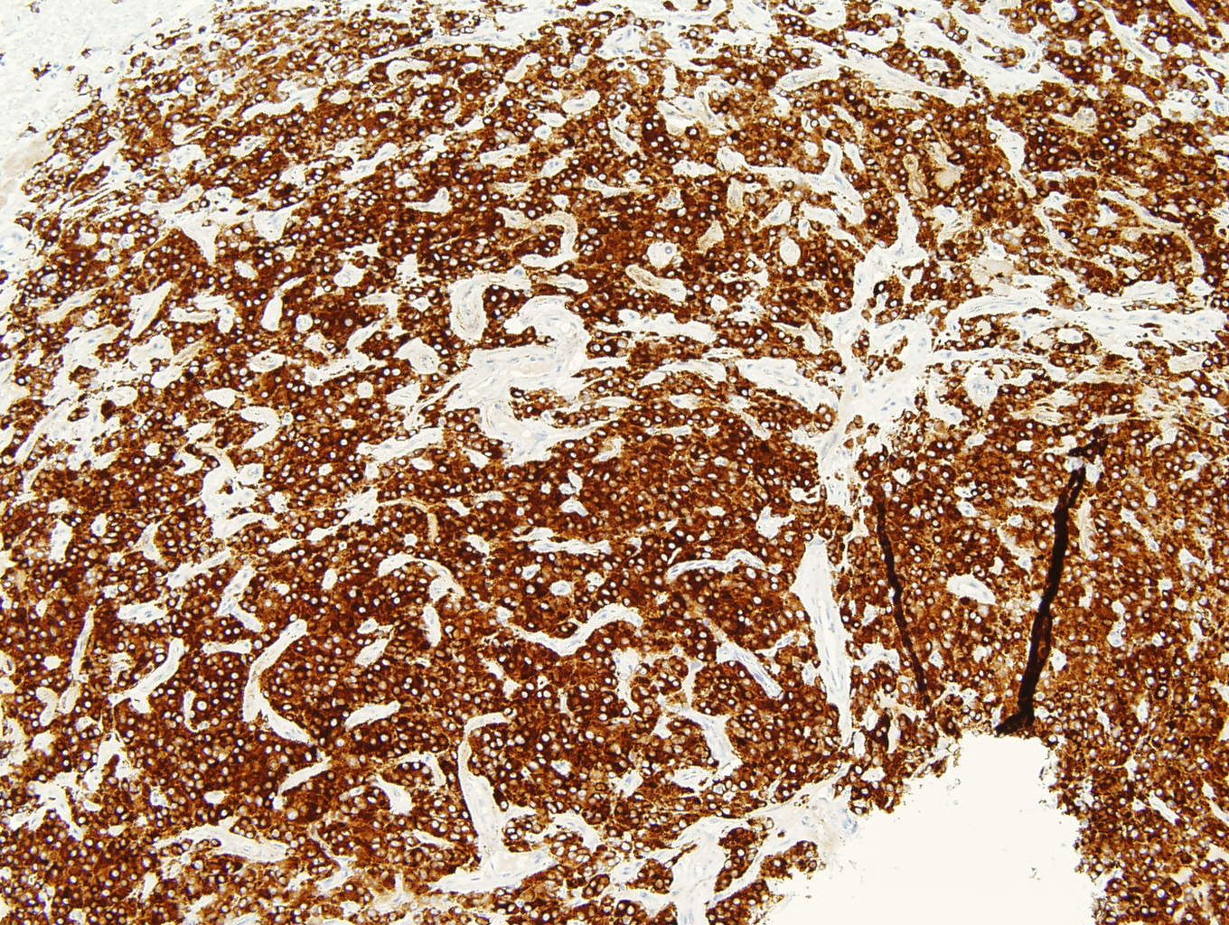
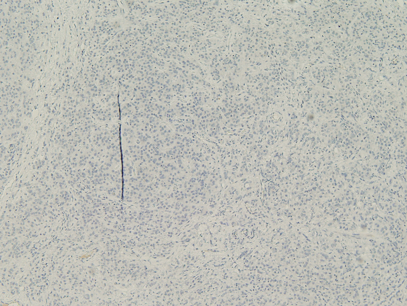
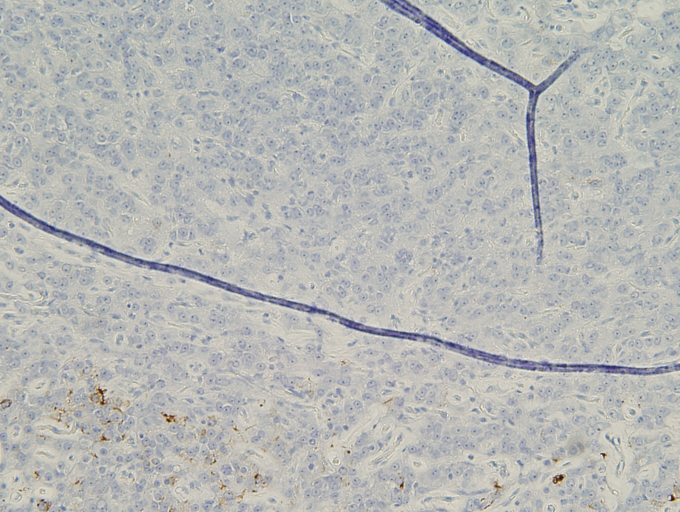
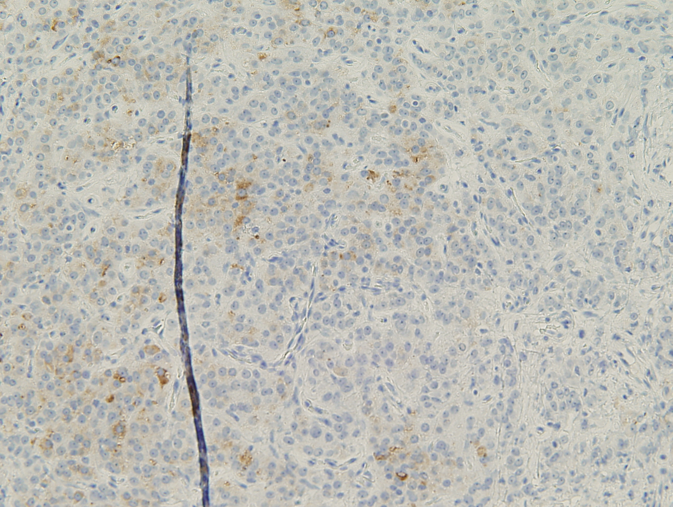
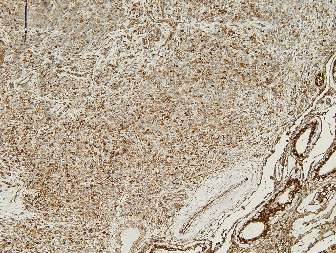
Immunihistochemistry
|
Figure 4. Desmin |
Figure 5. Inhibin |
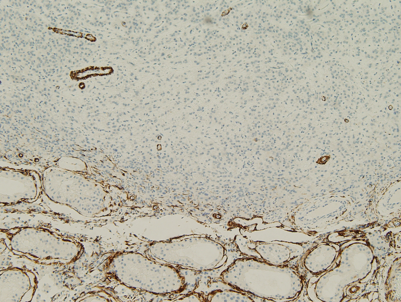 |
 |
|
Figure 6. CD30 |
Figure 7. CAM 5.2 |
 |
 |
|
Figure 8. Synaptophysin |
Figure 9. Vimentin |
 |
 |
 Meet our Residency Program Director