Residency Program - Case of the Month
June 2011 - Presented by Meighan Tomic, M.D.
Clinical history:
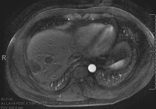
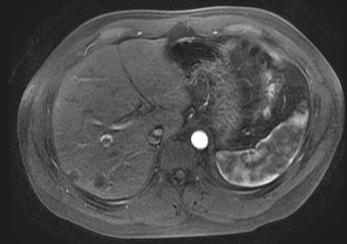
The patient is a 38 year old male, with a past medical history significant only for gastroesophageal reflux disease, who presented with burning epigastric pain. Right upper quadrant ultrasound revealed cholelithiasis and three, 2.0 – 2.7 cm, hypoechoic liver lesions. Follow up MRI showed multiple liver lesions with peripheral rim enhancement. Most lesions were peripheral, some lesions were confluent, and some sub capsular lesions had capsular retraction. PET scan was negative for other hyper metabolic lesions. Alpha fetal protein and CEA levels were not elevated. Hepatitis serologies were negative. Two CT guided core biopsies were obtained and were not diagnostic. Consequently a laparoscopic wedge resection was performed.
Abdominal MRI
 |
 |
Gross description:
Received fresh was an irregular, 2.0 x 2.5 x 1.0 cm, portion of liver with a 2.0 cm white sub capsular lesion.
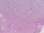
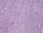
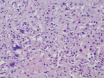
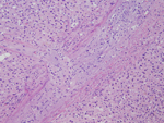
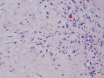
Microscopic photographs:
 |
 |
 |
 |
 |
Special Stains and Immunihistochemistry
|
Figure 1. Reticulin |
Figure 2. Factor VIII |
Figure 3. Factor VIII |
 |
 |
 |

 Meet our Residency Program Director
