
Residency Program - Case of the Month
July 2011 - Presented by Aram Millstein, M.D.
Clinical history:
The patient is a 54 year old woman with a history of melanoma of the right leg and a left adnexal mass that was found on CT when the patient presented to the ED with abdominal pain. The CT scan showed an adnexal mass with cystic and solid components and associated vascular flow, suspicious for an immature teratoma. The CA-125 was within the reference range. The patient followed up with Gynecologic Oncology and a bilateral salpingo-oophorectomy and total abdominal hysterectomy with staging was performed. Only the left ovary had significant pathology and is described below.
Gross description:
The left ovary was 5 cm in greatest dimension. It was sectioned to reveal a primary lesion with papillary orange-yellow and white cut surfaces. A cystic lesion was also present filled with soft cheesy material and hair. The histology is shown below.
Microscopic photographs:
|
Figure 1. Primary lesion, low power |
Figure 2. Primary lesion, medium power |
Figure 3. Primary lesion, high power |
Figure 4. Primary lesion, high power |
 |
 |
 |
 |
|
Figure 5. Primary lesion, high power |
Figure 6. Cystic lesion, low power |
Figure 7. Cystic lesion, low power |
Figure 8. Cystic lesion, low power |
 |
 |
 |
 |
Immunihistochemistry
|
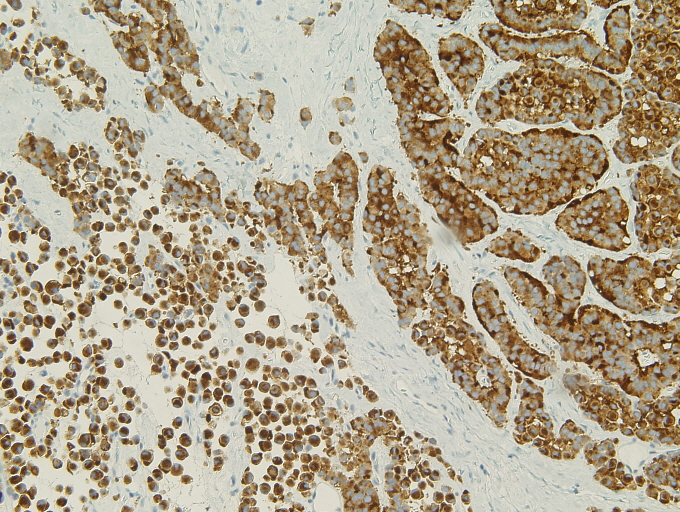
Fig 9. Synaptophysin |
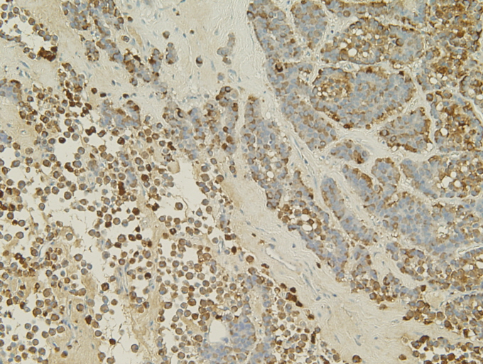
Fig 10. Chromogranin |
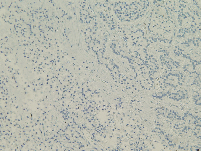
Fig 11. CD10 |
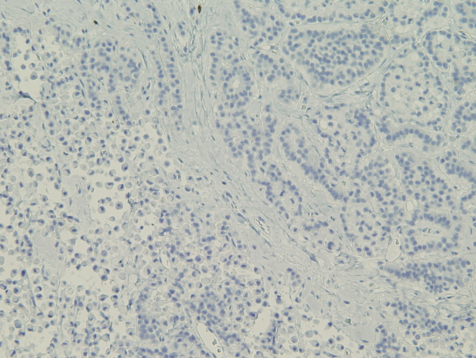
Fig 12. Calretinin |
 |
 |
 |
 |
 Meet our Residency Program Director