Residency Program - Case of the Month
March 2013 - Presented by John Rodrigo, M.D.
Clinical history:
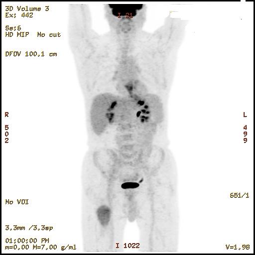
The patient is a 75 year old male who presented with dysphagia and epigastric discomfort. CT scans demonstrated a large distal esophageal mass and suspicious lymphadenopathy. An EGD at a later time confirmed this mass of the distal esophagus. Biopsy confirmed a high grade dysplasia. PET/CT around the same time demonstrated an enlarging intramuscular hypodense soft tissue mass in the right proximal thigh with interval development of internal dystrophic calcifications. The mass previously measured 2.8 x 2.8 cm but was now 5.6 x 4.4 cm.
PET scan:
Gross description:
Received was multiple cylindrical tan brown fragments from the right thigh soft tissue mass ranging in size from 1.2 cm in length by 0.1 cm in diameter to 1.5 cm in length by 0.1 cm in diameter with an aggregate of 0.9 x 0.3 x 0.2 cm.
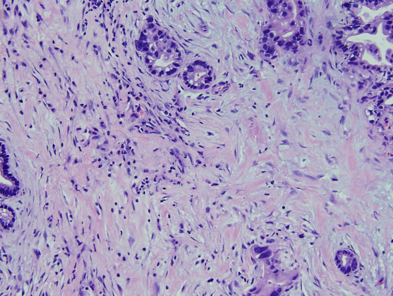
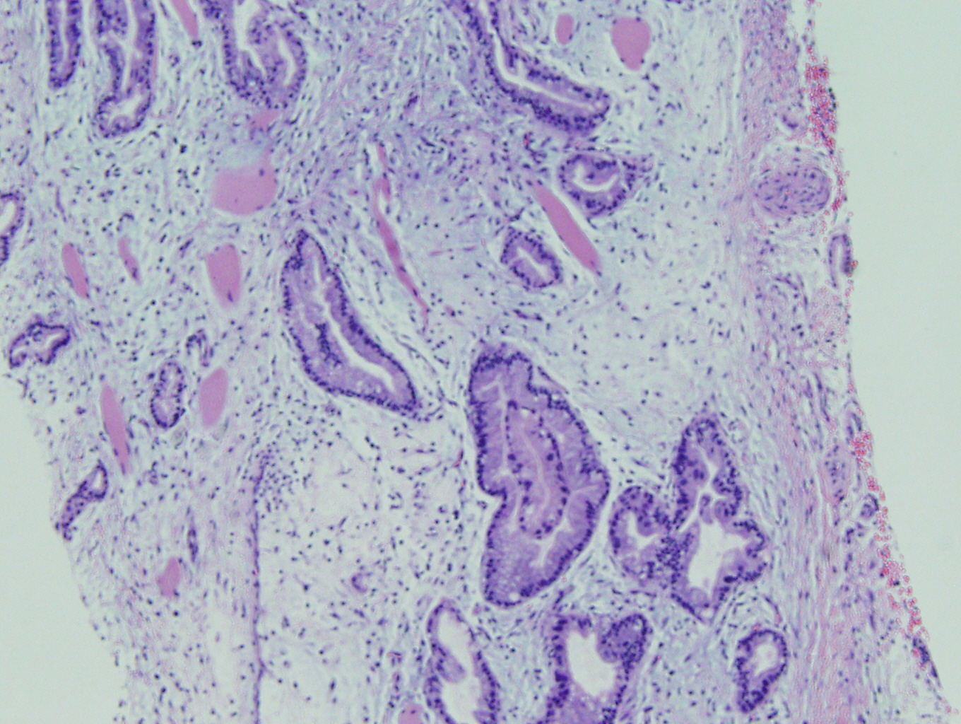
Microscopic pictures:
 |
 |
 |
|
 |
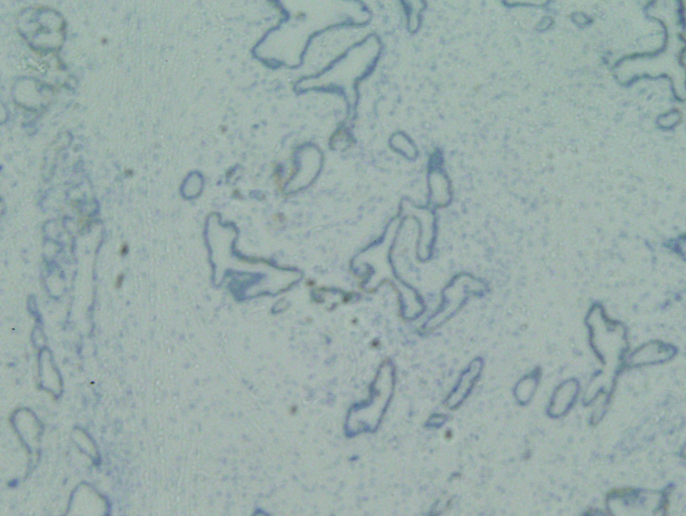 |
Immunohistochemistry stains:
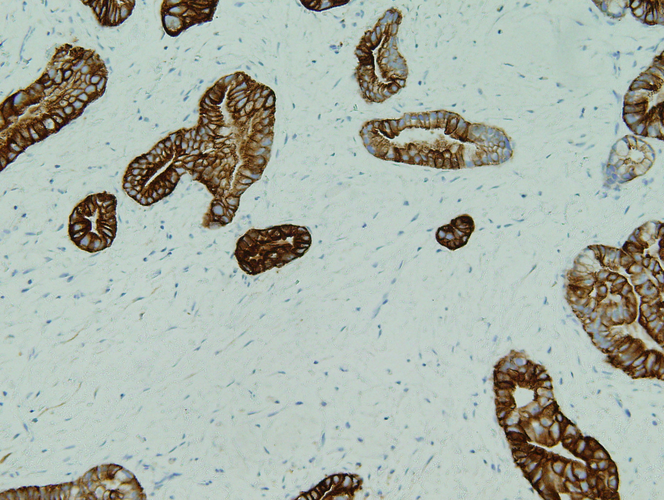
CK7 Positive (figure 1)
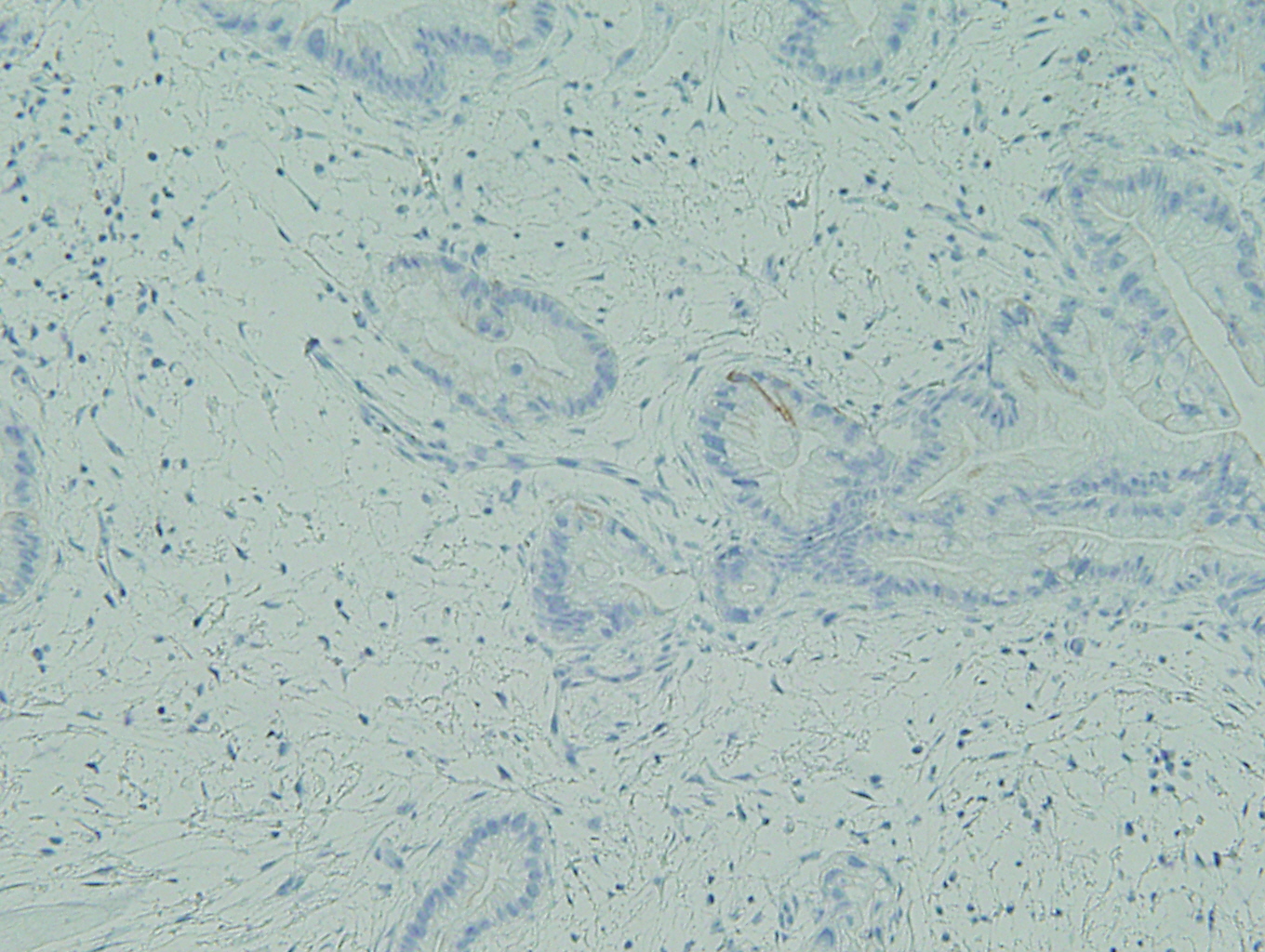
CK20 Negative (figure 2)
Her-2/neu 2+

 Meet our Residency Program Director
