Residency Program - Case of the Month
December 2013 - Presented by Shiloh Martin, M.D.
Clinical History:
The patient is a 50-year-old female with a past medical history of fibromyalgia, chronic pain, and 25 pack year smoking history, who presented to the ED at an outside hospital one year ago with severe left upper quadrant pain without fever, chills, anorexia, nausea, vomiting, diarrhea, constipation, or back pain. She additionally had a 40 lb weight loss (unintentional) over the prior 8 months. Ultrasound revealed pyelonephritis and a cystic lesion at the head of the pancreas. A subsequent CT confirmed a large abdominal mass (14.5 x 13.7 x 12.5 cm) with mixed cystic and solid features possibly arising from the head of the pancreas. An FNA and core biopsy were performed at the outside hospital (reviewed by UCD Pathology) and demonstrated a neoplasm consistent with (answer). She was referred to UCD GI surgery, and the patient decided to undergo medical management at that time with Gleevec (imatinib). Several follow up CT scans demonstrated a large mass arising from the antrum of the stomach, which was stable over time. However, over the next several months, the patient was unresponsive to imatinib and her symptoms worsened (increasing anorexia, nausea, vomiting, fatigue) and she elected to proceed with surgical resection. Intraoperative findings were as follows: large mass arising from the distal antrum without invasion of other structures, no evidence of pancreatic involvement, no evidence of gross metastatic disease in the abdomen. Distal gastrectomy was performed.
Gross Description:
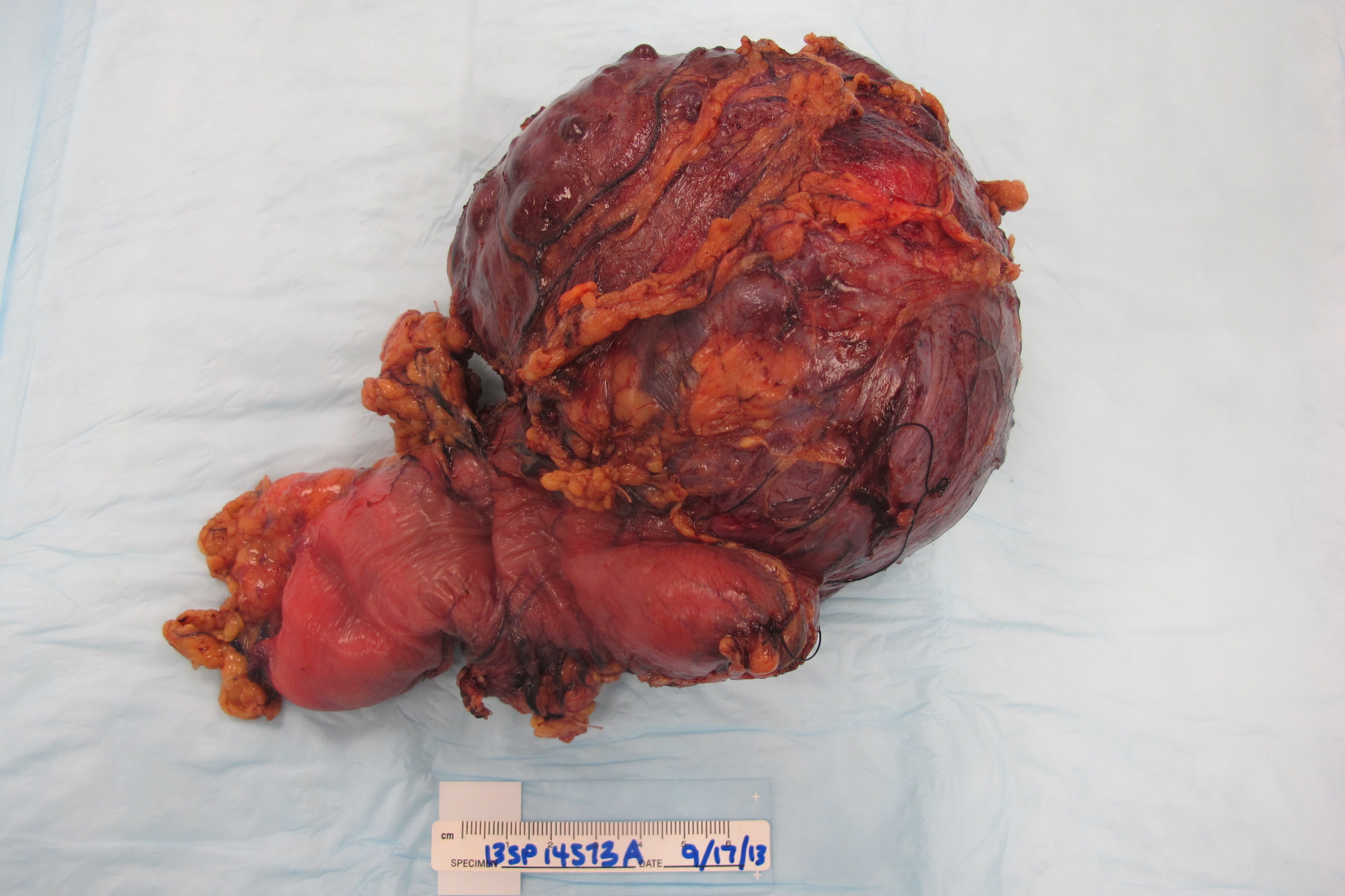
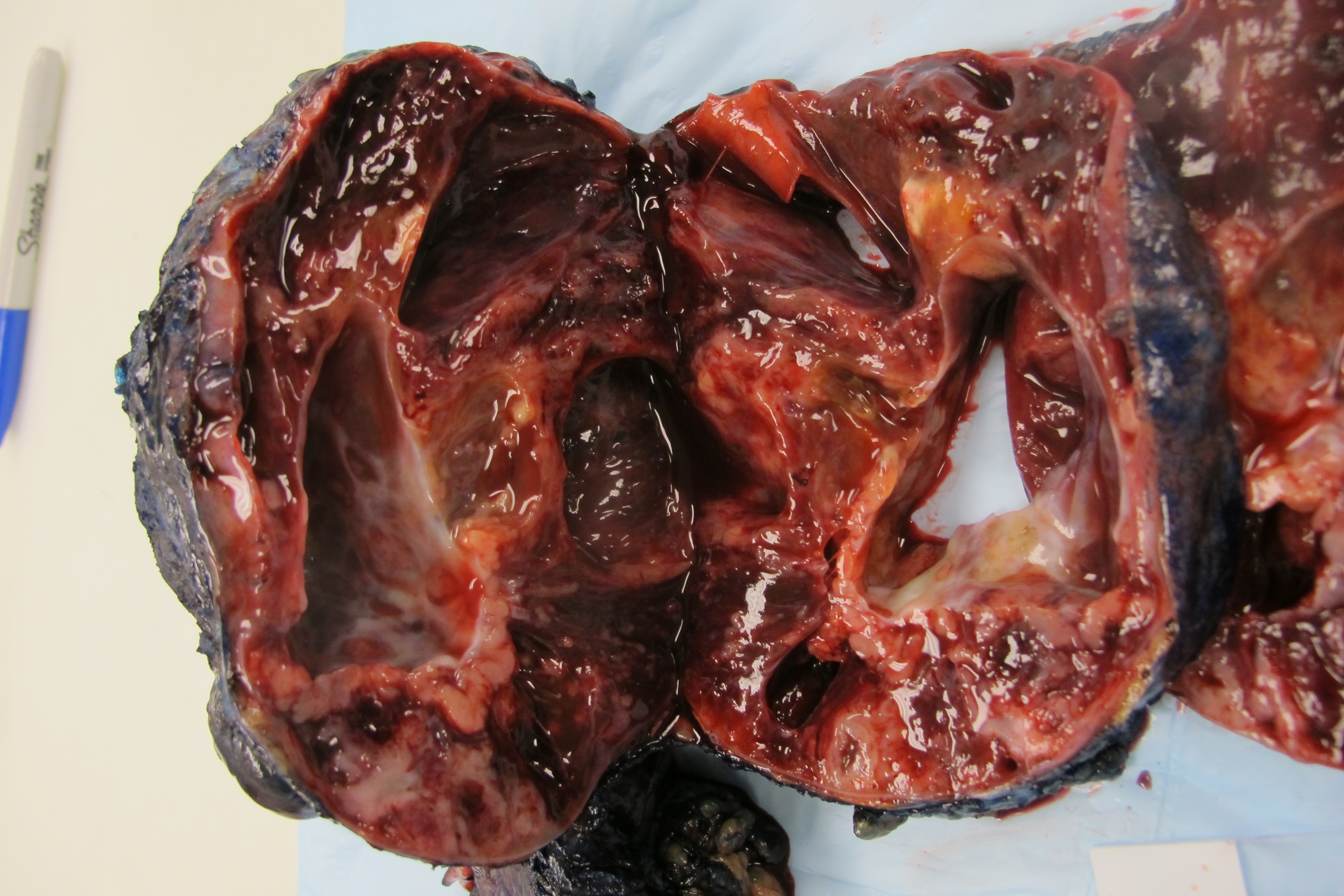
Received is an oriented gastroduodenectomy specimen consisting of a wedge of antrum and portion of duodenum (15 x 6 x 3.5 cm) with a large, round, multicystic, purple-grey mass (14 x 13 x 10 cm) consisting of multiple blood and serous filled cysts ranging in size from 1.0 cm to 5.5 cm. The cysts are lined by grey-white 0.1 cm thick walls without solid excrescences. Between the cysts are solid areas consisting of tan-white and red gelatinous surfaces. The mass is well encapsulated and grossly appears to arise from the submucosa at the muscularis propria along the greater curvature of the stomach. The mass measures 2.5 cm to the duodenal margin and 4.5 cm to the nearest gastric margin. The rugated pink-tan gastric mucosa is uninvolved and without additional masses, ulcers, or polyps. Along the greater curvature is a portion of yellow mesenteric fat (2.5 x 3.5 x 2.0 cm) containing multiple lymph nodes. Inking scheme: black - duodenal margin; purple - gastric margin; blue - mass.
 |
 |
Micro Description:
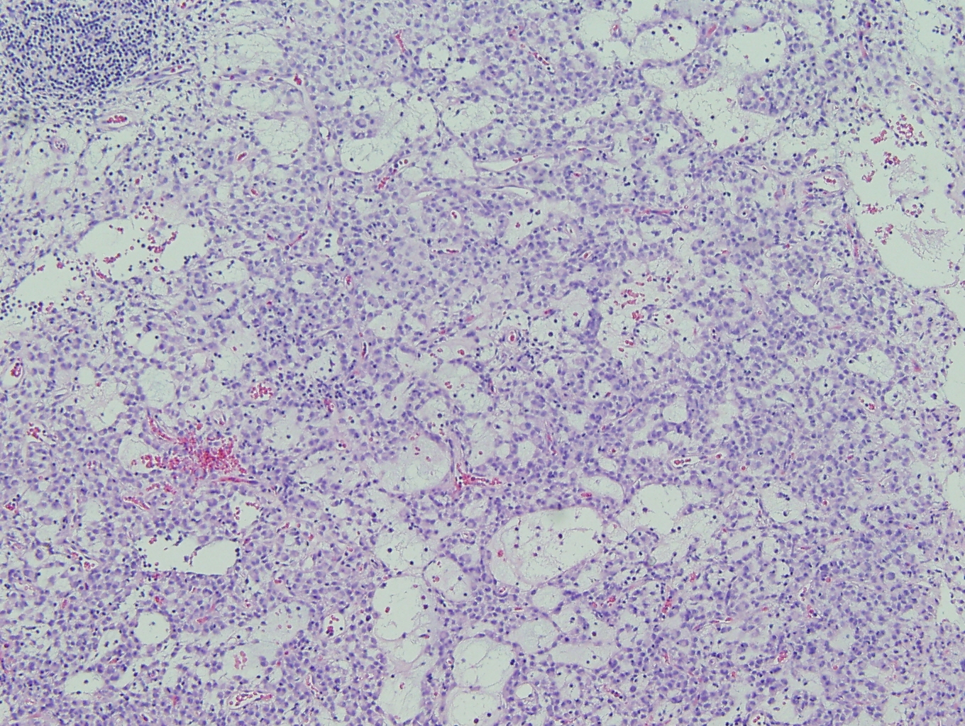
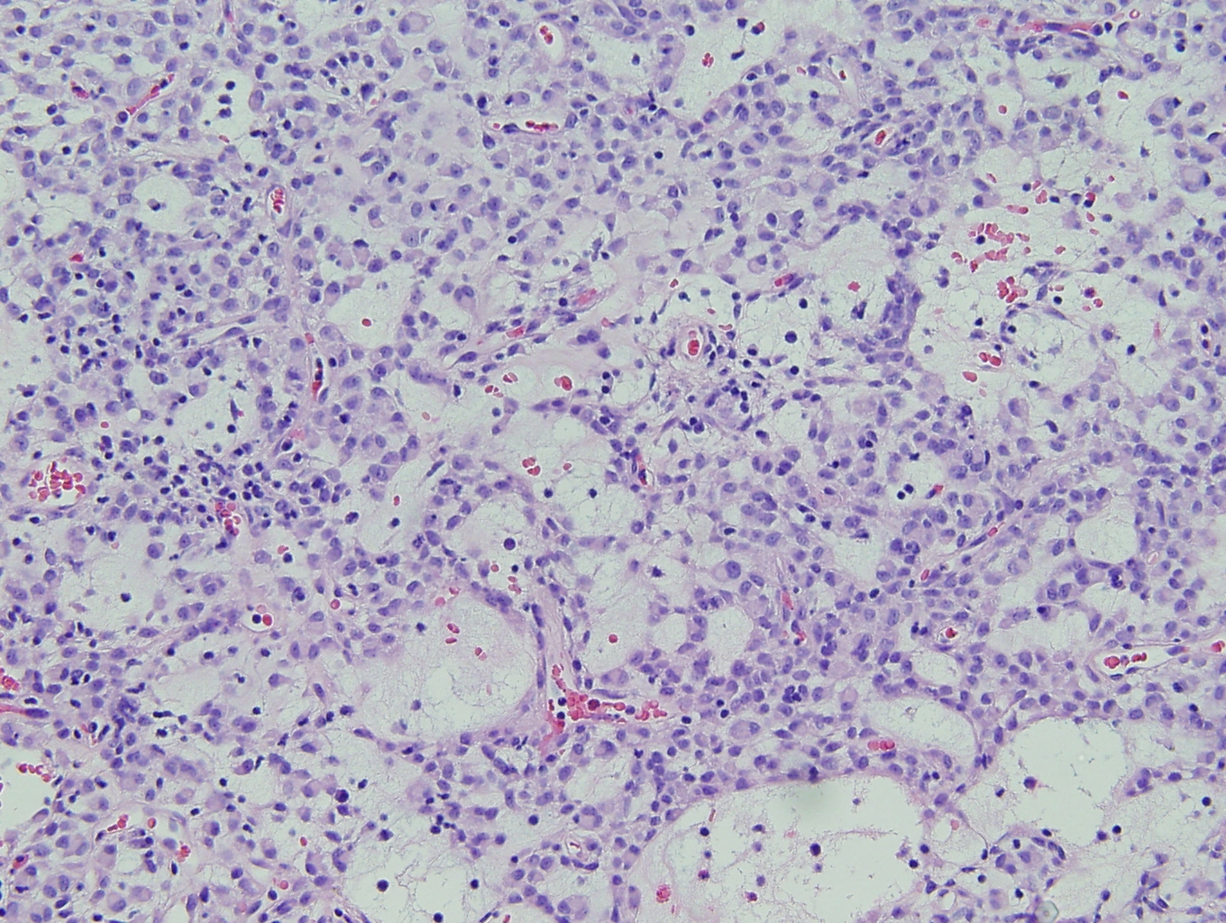
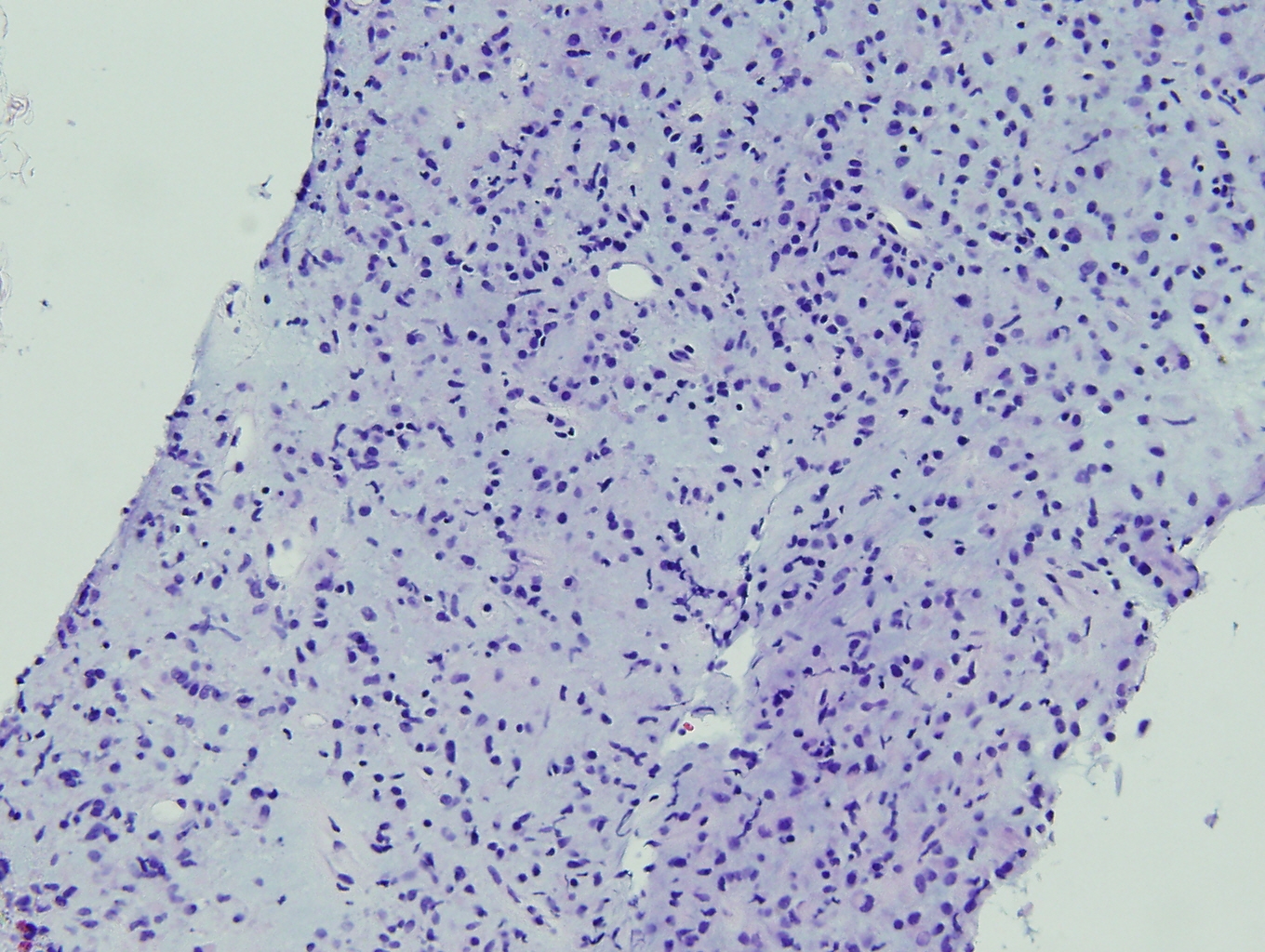
Predominantly sheets of large epitheloid cells with eosinophilic cytoplasm with mucopolysaccharide droplets or cytoplasmic clearing. Nuclei are round to oval with small nucleoli with occasional binucleation.
 |
 |
| Stomach Tumor | Stomach Tumor |
 |
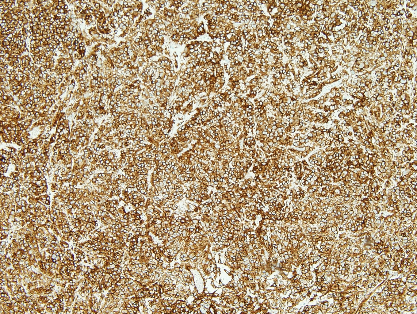 |
| Pancreatic Biopsy | Dog-1 IHC |
Immunohistochemistry:
Gastroduodenectomy specimen:
| Dog-1 | positive |
| Desmin | focally positive |
| Ki67 |
positive (5%) |
| C-KIT (CD117) | negative |
| CD31 | negative |
| CD34 | negative |
| CD68 | negative |
| S100 | negative |
| AE1/AE3 | negative |
| HMB45 | negative |
| SMA | negative |
Pancreatic biopsy (IHC reviewed at UCD):
| Dog-1 | positive |
| Actin | focally positive |
| Desmin | negative |
| C-KIT (CD117) | negative |
| CD34 | negative |
| S100 | negative |
| Cytokeratin | negative |

 Meet our Residency Program Director
