Residency Program - Case of the Month
February 2014 - Presented by Christina Di Loreto, M.D.
Clinical History:
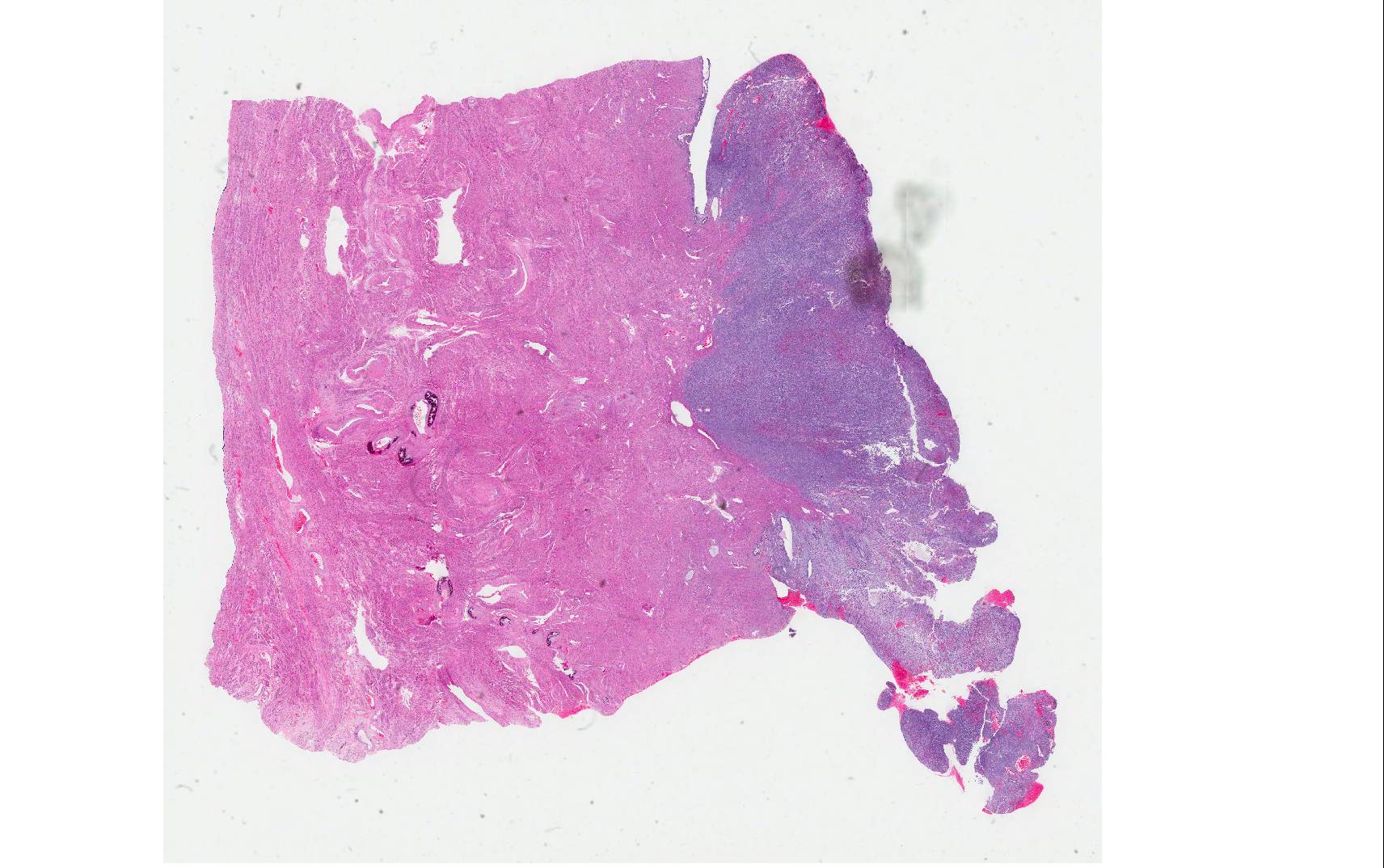
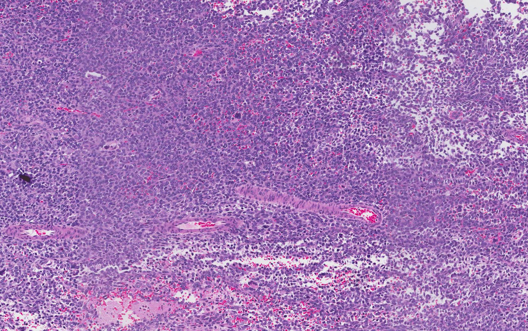
The patient, a 91 year old female, presented with a 6 month history of vaginal bleeding and mild abdominal discomfort. CT scan demonstrated a 2.6 x 3.2 x 5.1 cm mass in the uterus, an 18 mm right pelvic sidewall lymph node, and a 19 mm left adrenal mass. Biopsy of the endometrium performed at an outside institution was interpreted as high-grade undifferentiated malignant neoplasm, favoring undifferentiated uterine sarcoma. A total laparoscopic hysterectomy and bilateral salpingo-oophorectomy was performed.
Gross Description:
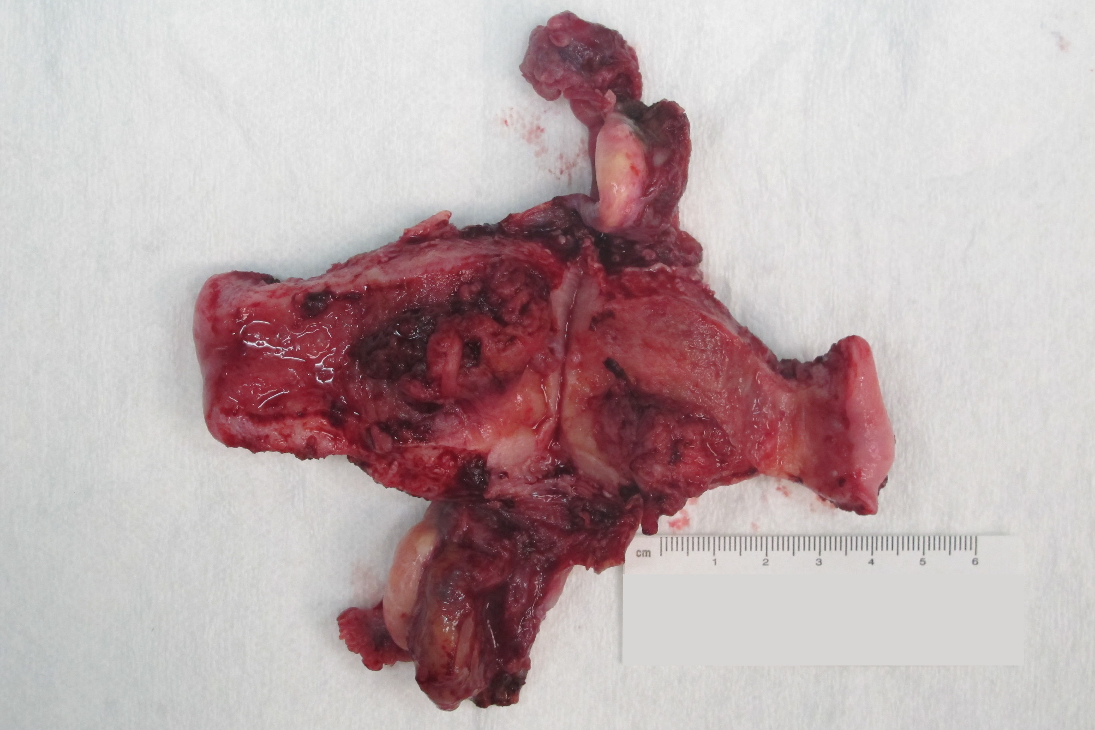
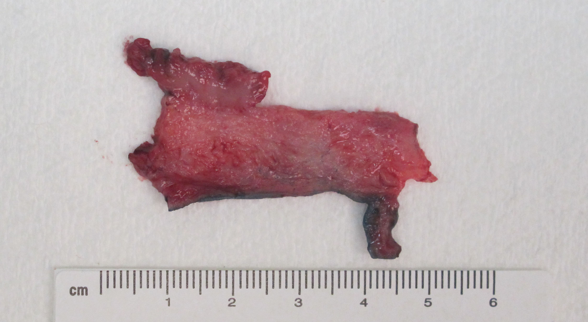
Received was a 78.1 gram uterus with cervix and attached bilateral adenexa. Two superficial, broad-based, polypoid masses arising from the anterior and posterior endometrium were identified, measuring 3.0 x 1.5 x 0.7 cm and 3.6 x 2.1 x 1.0 cm, respectively.
Gross Images:
 |
 |
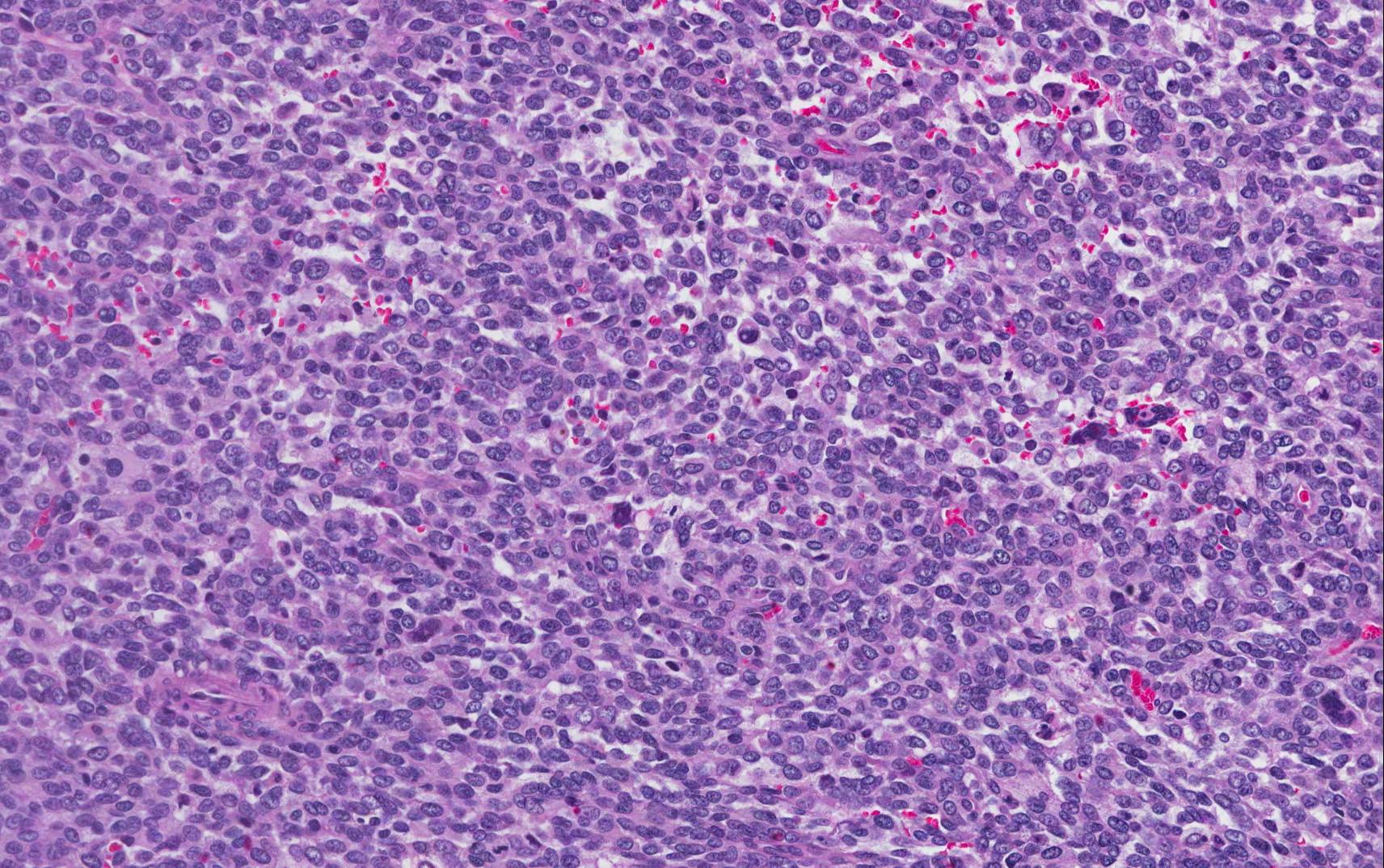
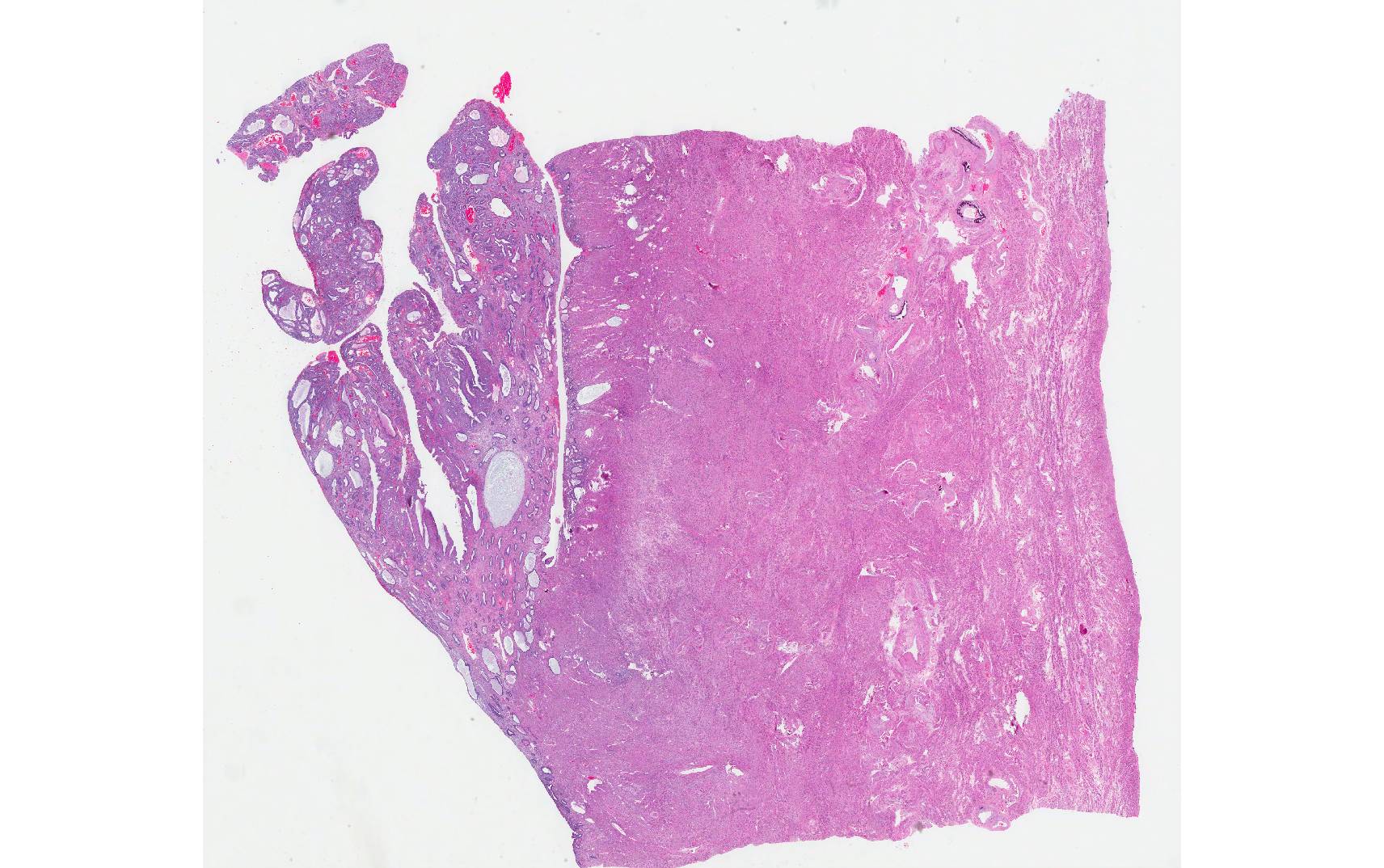
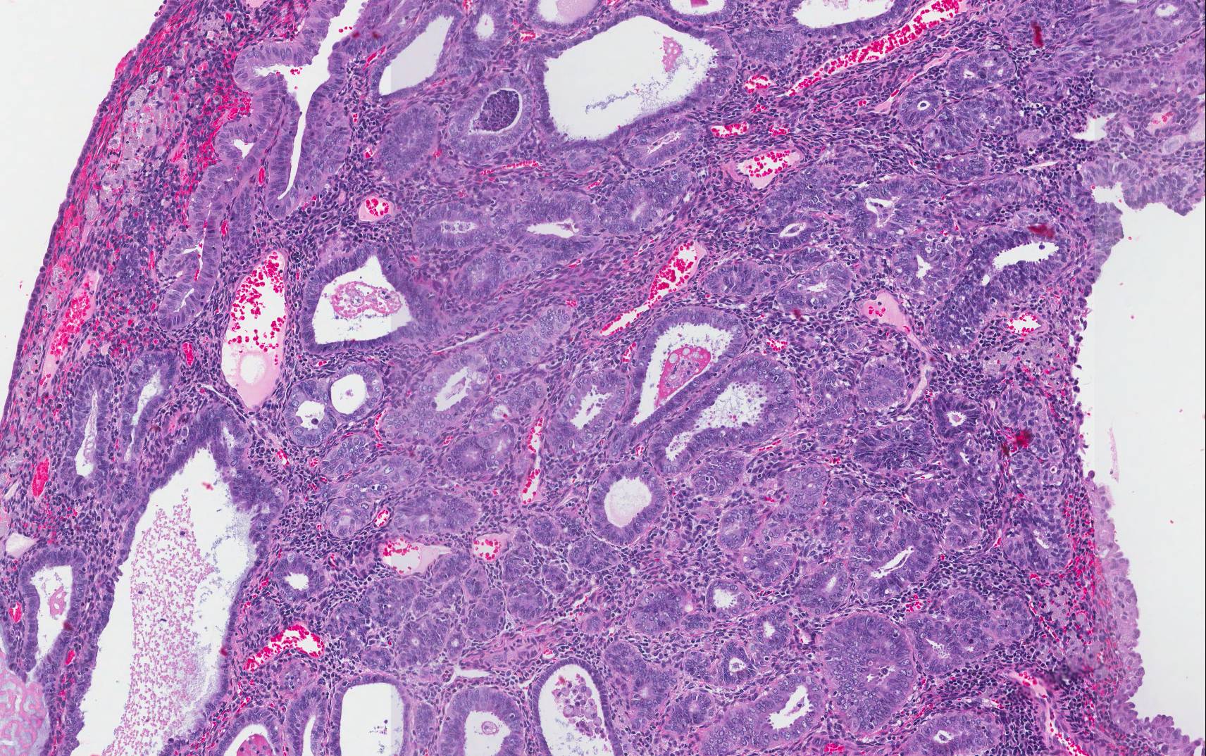
Microscopic Images:
 |
 |
 |
||
|
Image 1 |
|
Image 2 |
|
Image 3 |
 |
 |
|||
| Image 4 | Image 5 |
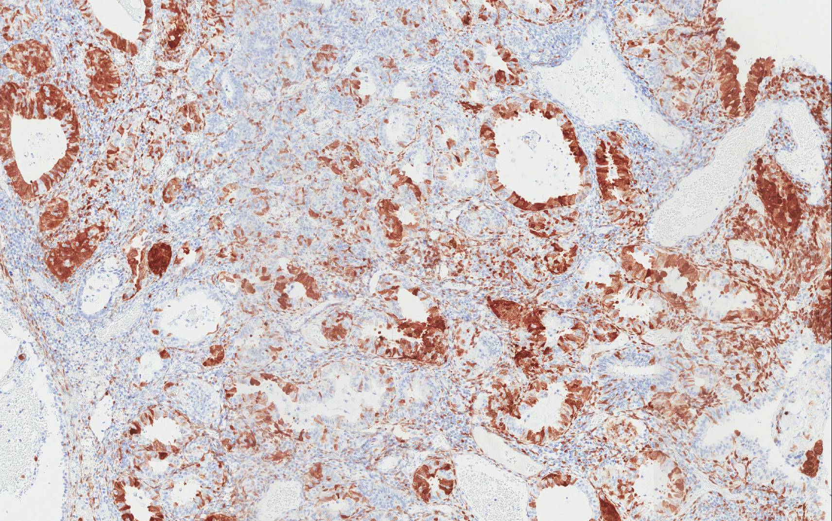
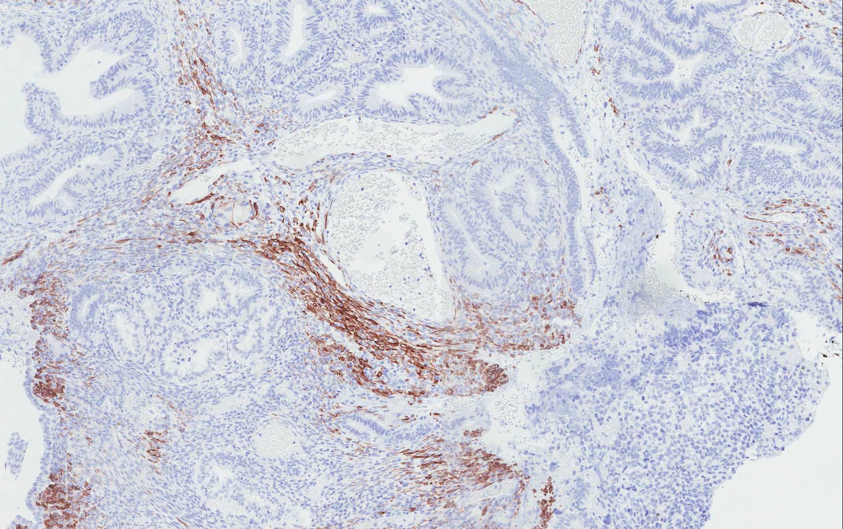
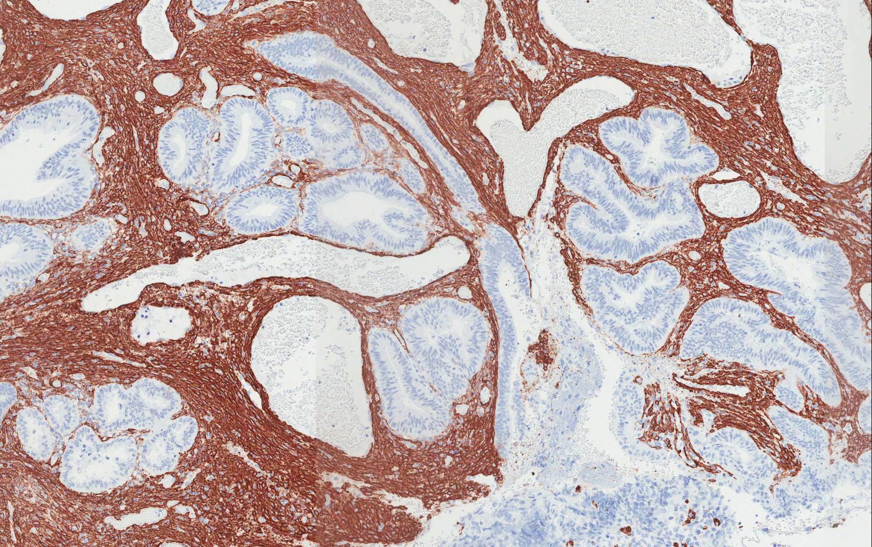
Immunohistochemistry:
 |
 |
|
| AE1/AE3 | p16 | |
 |
 |
|
| Desmin | SMA |
P53: Negative
CD10: Negative

 Meet our Residency Program Director
