Residency Program - Case of the Month
April 2015 - Presented by Dr. Elham Vali Khojeini
History:
A 66-year-old female with a past medical history of peptic ulcer disease presents to the Emergency Department with 5 weeks of vague epigastric abdominal pain with non-bloody non-bilious emesis. She does not report any weight loss, fever, chills or lymphadenopathy.
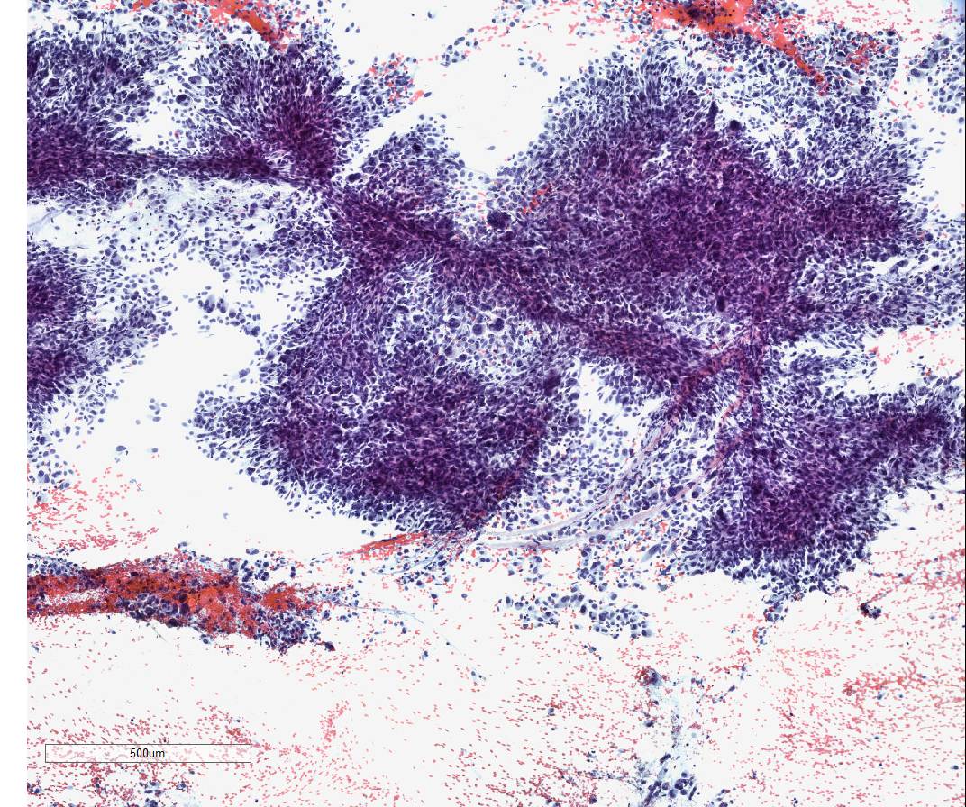
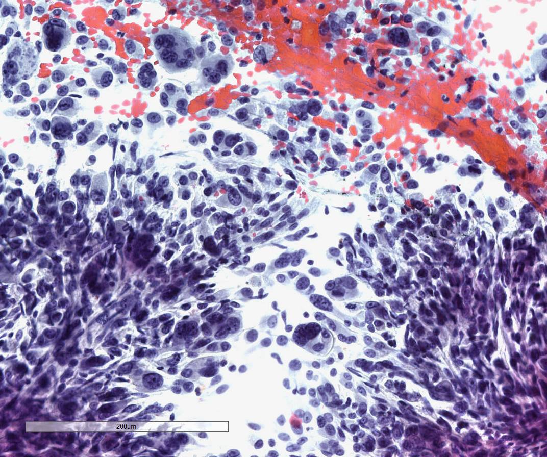
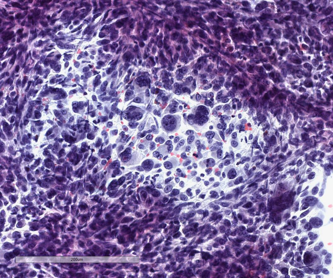
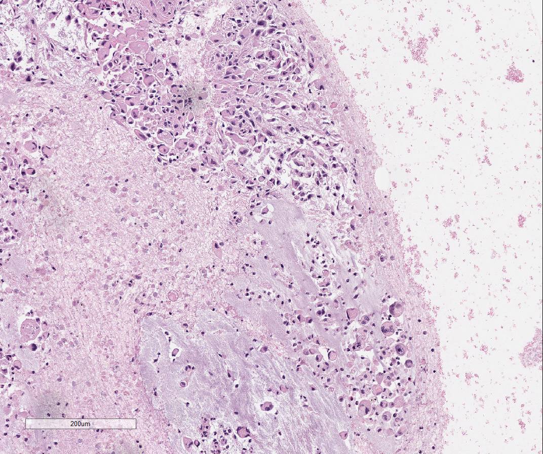
A week before, she was seen at an outside hospital with a chief complaint of abdominal pain and was given opioids and anti-reflux medicine, which did not improve her symptoms. On her workup, labs were unremarkable, however, a CT scan demonstrated a large heterogeneous mass (7.2 x 6.3 x 6.6 cm) arising from the pancreatic body with chronic occlusion of the splenic vein. The mass has a large cystic component which may be related to an associated pseudocyst or necrosis. Also, there are subcentimeter liver lesions. An ultrasound/doppler-guided fine needle aspiration was performed which showed the following images:
Answer


 Meet our Residency Program Director
