Residency Program - Case of the Month
June 2015 - Presented by Dr. Adam Stelling
History
The patient is an 82 year old man who presented with fever, diaphoresis, fatigue, and dysphagia. On exam, he was noted to have prominent lymphadenopathy including enlarged tonsils as well as splenomegaly. He had been seen previously for the tonsillar enlargement with a biopsy showing necrotizing atypical lymphoid infiltrate with increased eosinophils. At this time, a right axillary lymph node excisional biopsy was performed for definitive characterization of the nodal pathology.
Microscopic Description
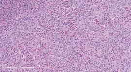
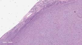
Sections from the lymph node show complete effacement of normal architecture by a diffuse proliferation of sheets and fascicles of monocytoid spindle to ovoid cells with moderate eosinophilic cytoplasm and slightly enlarged irregular grooved nuclei. There are frequent mitoses and abundant eosinophils in the background.
 |
 |
|
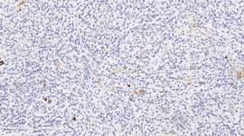
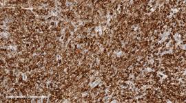
| CD1a | CD4 | |
 |
 |
|
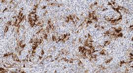
| CD23 | High Power H&E | |
 |
 |
|
| Low Power H&E | S100 |
Sample Morphologic Differential Diagnosis
- Langerhans cell histiocytosis
- Follicular dendritic cell sarcoma
- B and T Large cell lymphomas
- Classical Hodgkin lymphoma, lymphocyte depleted type
- Kimura lymphadenopathy
Immunohistochemistry
CD23: Positive
CD68: Positive
CD1a: Negative
S100: Negative
CD4: Positive
Ki67: Positive in many of the atypical cells
CD30: Negative
CD15: Scattered positive eosinophils
CD20: Scattered positive lymphocytes
CD3: Scattered positive lymphocytes
Fascin: Positive

 Meet our Residency Program Director
