Residency Program - Case of the Month
September 2015 - Presented by Dr. Jessica Rogers
History:
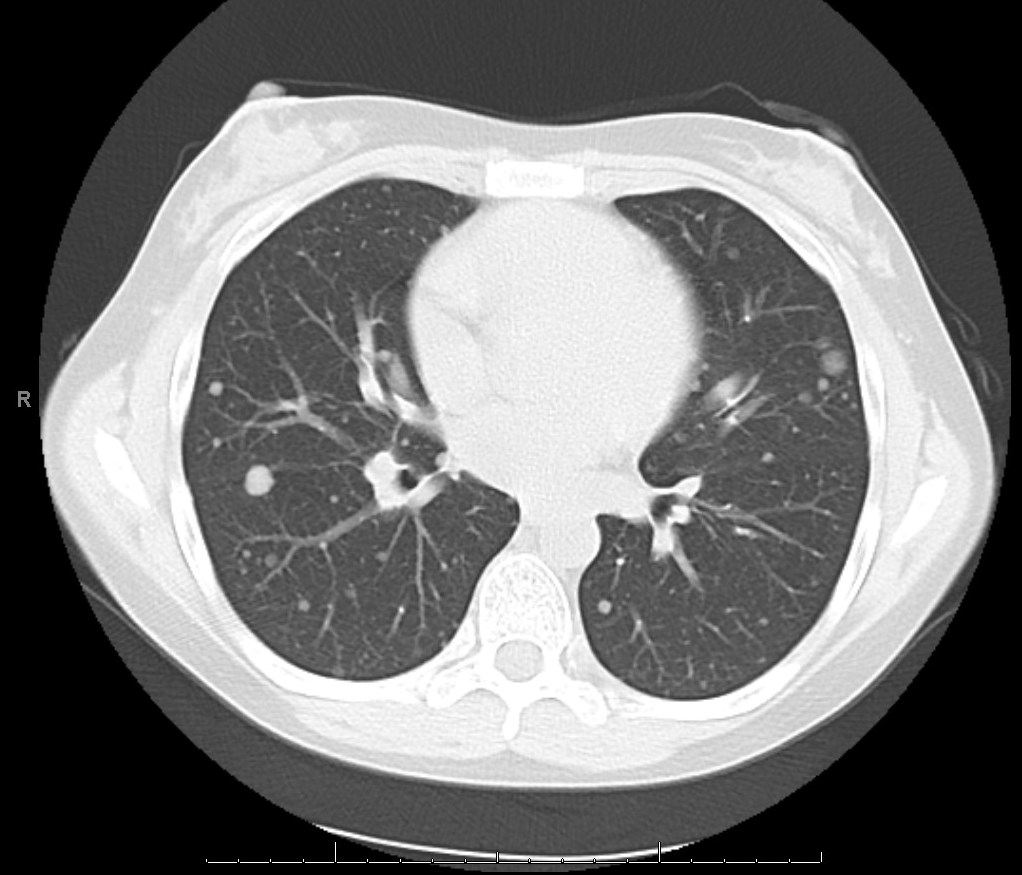
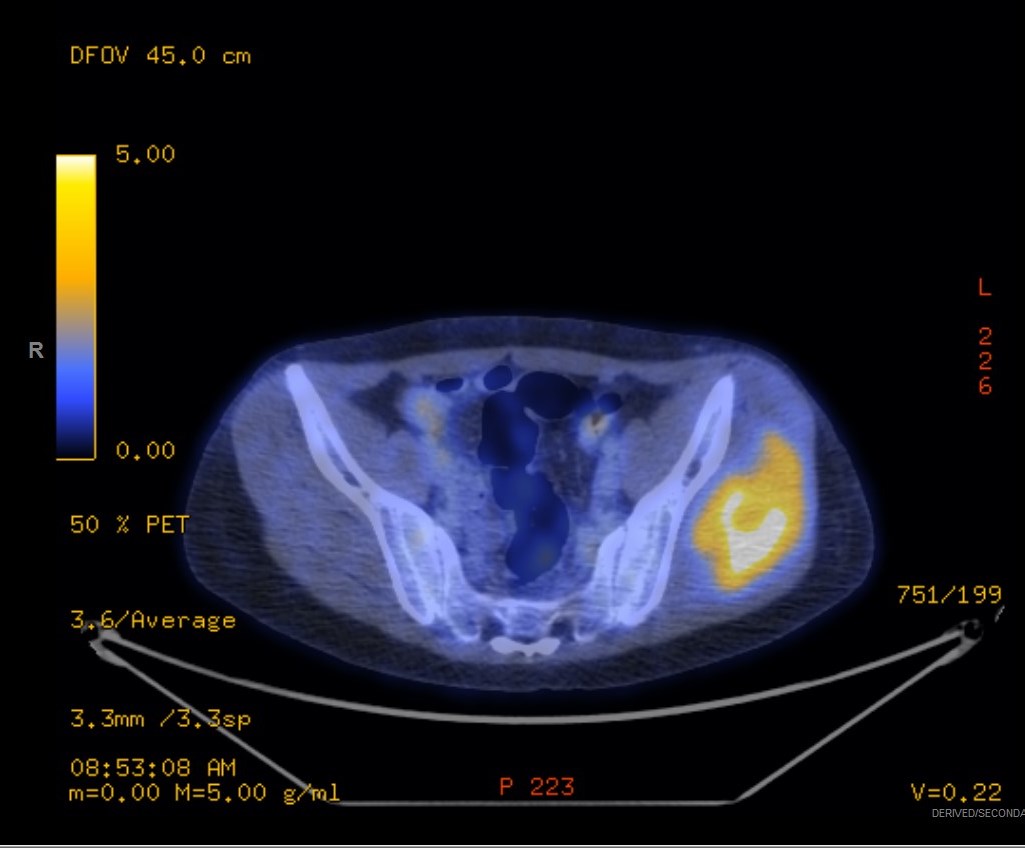
A 20-year-old female with history of Systemic Lupus Erythematosus (SLE) and lupus nephritis causing renal failure for which she is on hemodialysis. She was on chronic immunosuppression and subsequently developed a left thigh nodule and numerous lung nodules. Incisional biopsy of the left anterior thigh mass showed suppurative granulomatous dermatitis containing fungi consistent with chromomycosis and the patient was started on Itraconazole. The pulmonary nodules (Figure 1) did not change in size with anti-fungal treatment, so concern was raised regarding their malignant potential. Ultimately a whole body PET/CT was performed in search of a primary malignancy, which showed a FDG avid left gluteal tumor (Figure 2). A CT guided core needle biopsy of the mass was obtained.
| Figure 1 | Figure 2 | |
 |
 |
Gross:
Received were eight cores ranging from 0.4 cm to 2.8 cm in length and measuring 0.1 cm in diameter. The specimen was entirely submitted.
Histology:
The architecture is composed of nests of uniform eosinophilic cells in an alveolar pattern. The cells have abundant granular eosinophilic cytoplasm with some cleared out cells. The nuclei are large, round, and have single prominent nucleoli (Figure 3). Mitotic figures are scarce.
Immunohistochemistry
s100:
negative
Melan A:
negative
HMB-45:
negative
AE1/3:
negative
EMA:
negative
SMA:
negative
Desmin:
negative
Ki67:
increased (8-10% positive)
CD138:
negative
TEF3:
positive (Figure 4)
Myogenin:
negative
| Figure 3 | Figure 4 | |
 |
 |
Answer

 Meet our Residency Program Director
