
Residency Program - Case of the Month
August 2017 - Presented by Nicholas Coley (Mentored by Regina Gandour-Edwards)
Clinical History
The patient is a newborn baby boy delivered at 37 weeks with a large 9 cm multicystic mass overlying his neck that significantly impaired his respiratory efforts. The mass was removed by ENT and at the time of surgery, frozen sections demonstrated tissue consistent with thyroid.
Microscopic Description
The tumor demonstrates a multicystic structure with numerous heterologous elements. Squamous epithelium/mucosa, cartilage, bone, adnexal structures, and columnar epithelium resembling respiratory epithelium are all present. There are also three regions present at low power that demonstrate elongated cells with an increased nuclear to cytoplasmic ratio arranged in rosettes around a central lumen (Figure 1). The nuclei of these cells are elongated and hyperchromatic with regular nuclear membranes. The cells stain positively for synaptophysin (Figure 2). Increased mitotic/proliferative activity (Figure 3) and single cell necrosis are also present in these regions. Also present are several cystic structures lined with a cuboidal, darkly pigmented single layer of cells on one aspect that abruptly transitions into a lining of elongated cells several cell layers thick that are perpendicularly oriented to the lumen (Figure 4).
Click on image to enlarge.
Figure 1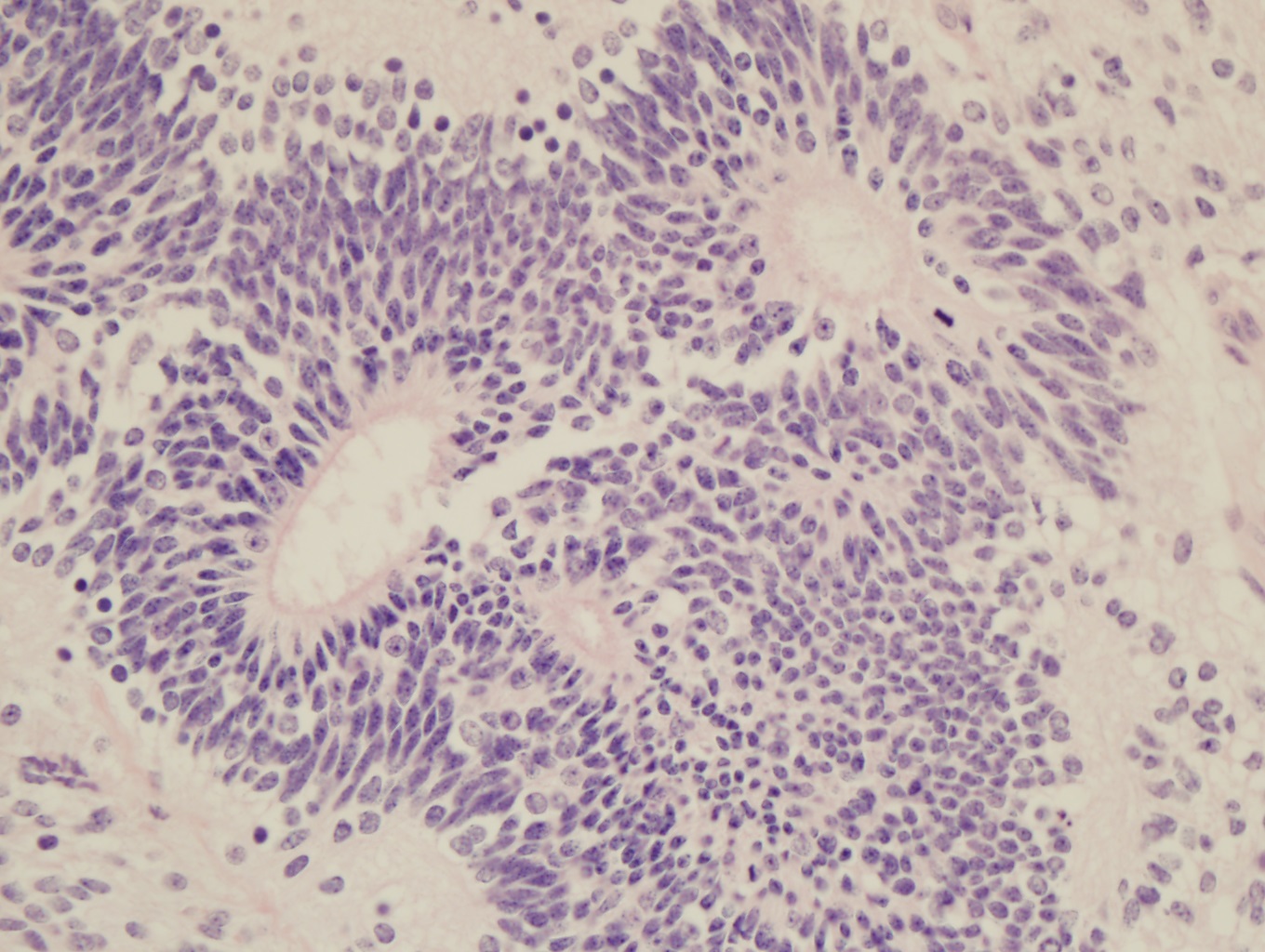
Figure 2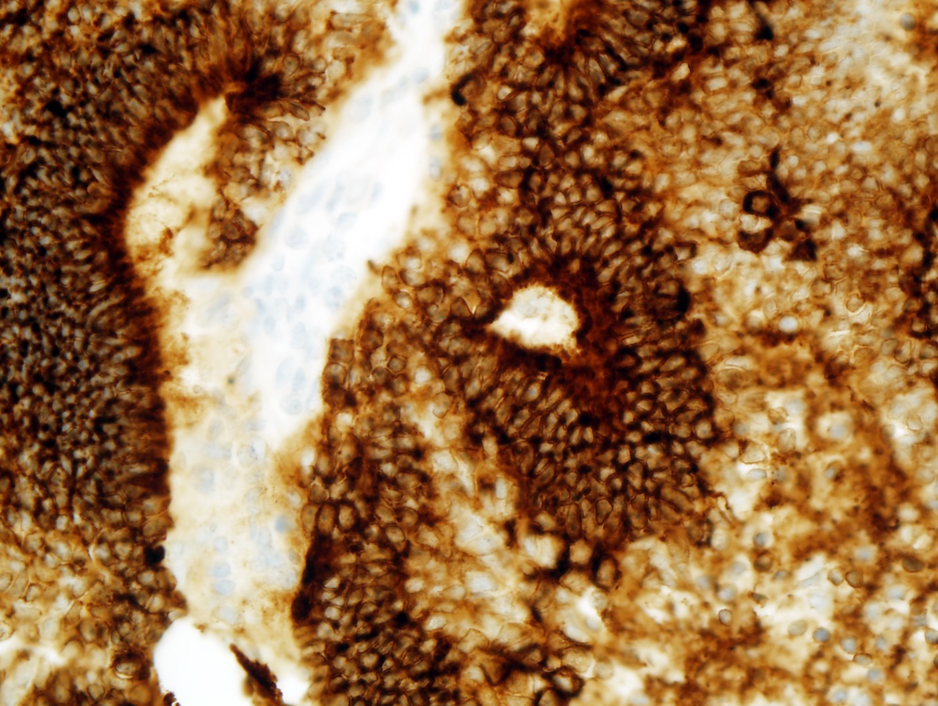
Figure 3
Figure 4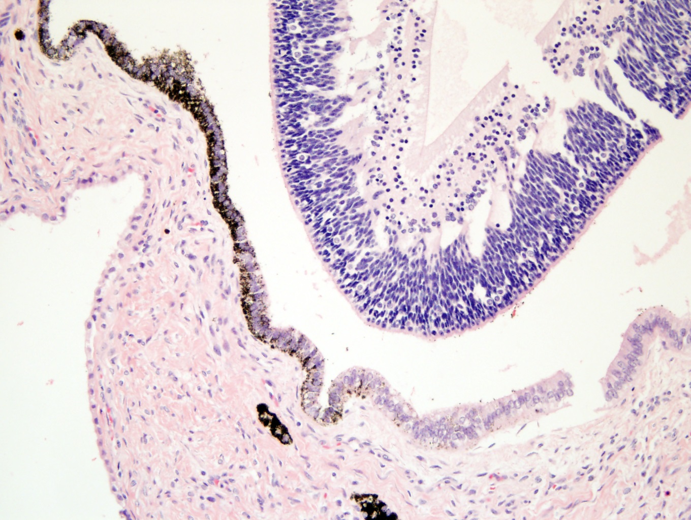
What is the diagnosis?
Choose one answer and submit.
 Meet our Residency Program Director