
Residency Program - Case of the Month
February 2018 - Presented by Guofeng Gao (Mentored by: Chihong Zhou)
Clinical History
A 78-year-old man with a history of biliary colic admitted to outside hospital in for gallstone pancreatitis. He underwent laparoscopy for gallbladder removal but the procedure was aborted due intraoperative findings of a cirrhotic liver. The follow up CT scan showed a hypodense lesion in the pancreas and a follow up EUS with biopsy demonstrated a mass in the uncinate process. Endoscopy ultrasound guided transduodenal fine needle aspiration of the pancreas mass was performed.
Microscopic Description
Both smears by Diff-Quik stain and by Papanicolaou stain and the cell block are hypercellular and show large cells as crowded clusters, syncytial groups and singly dispersed, which are composed of likely three different types of cells. First are the osteoclastic giant cells. They have multiple centrally located and bland looking nuclei, nuclear overlap, smooth nuclear membranes, pale chromatin, distinct and small nucleoli and moderate cytoplasm. Second are the pleomorphic giant cells. They are large in size and demonstrate markedly enlarged, often bilobated to multilobated, binuclear to multinuclear, highly pleomorphic and often hyperchromatic nuclei, some with coarse chromatin, very irregular nuclear contours, occasional nucleoli and sometimes abnormal mitoses. The cytoplasm varies from small to moderate to abundant in amount. Third type of cells is not easy to identify. When carefully search, there are some spindle shaped tumor cells and some atypical histiocytic looking tumor cells. They have slightly enlarged round to oval nuclei, smooth to irregular nuclear membranes, small indistinct nuclei and moderate to abundant cytoplasm.
Immunohistochemistry (IHC) Results
| CK AE1/AE3 | Positive (Fig. 8) | |
| GLYPICAN 3 | Negative | |
| HEP PAR1 | Negative | |
| CK7 | Very Foally Positive | |
| CK20 | Negative | |
| CA 19-9 | Negative | |
| SMAD4 | Negative | |
| CD56 | Negative | |
| p-53 | Positive (Fig. 9) | |
| CD68 | Negative (Fig. 10, positive in osteoclast-like giant cells) |
Click on image to enlarge.
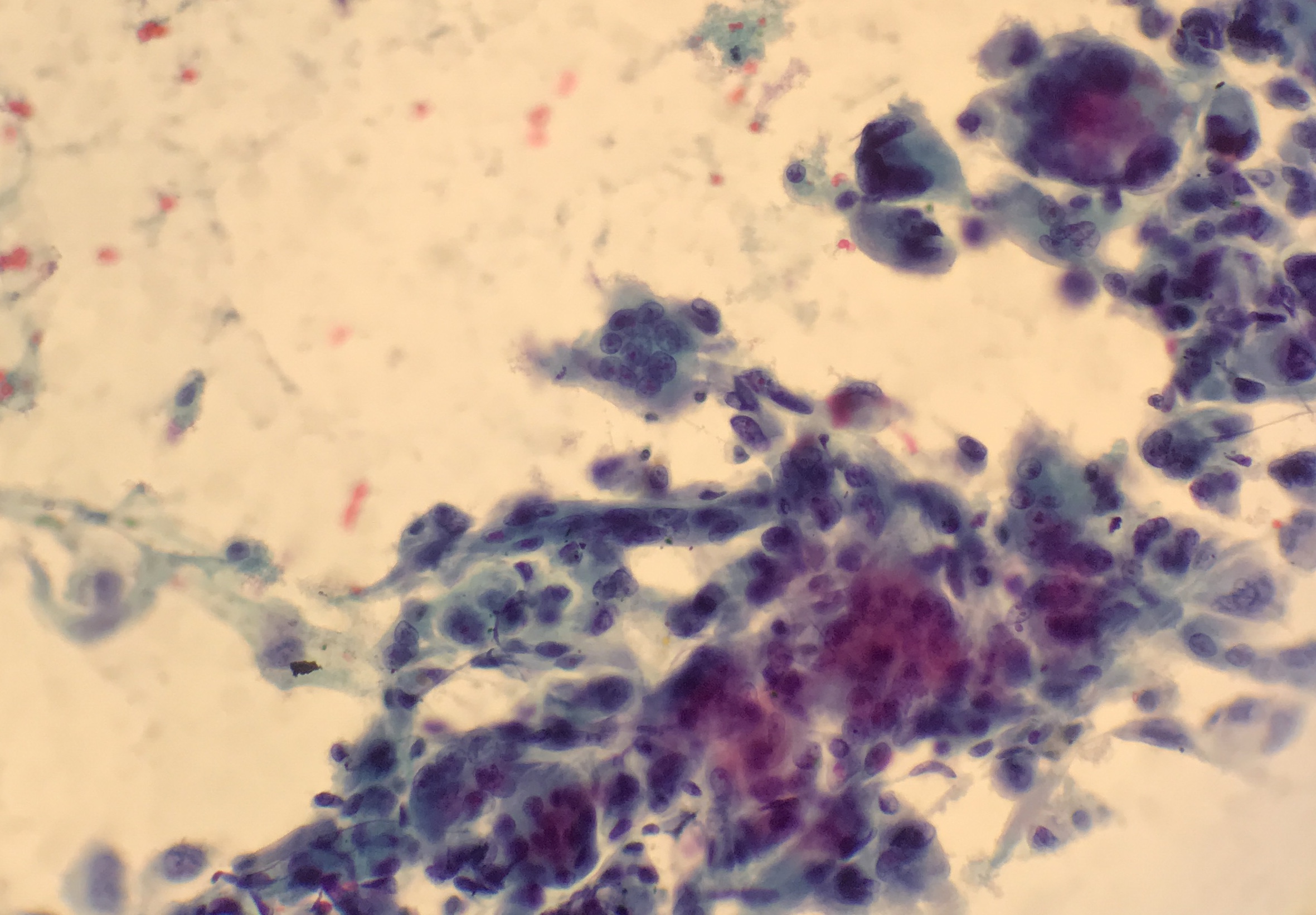
Figure 1: FNA Smear by Diff-Quik stain (400x)
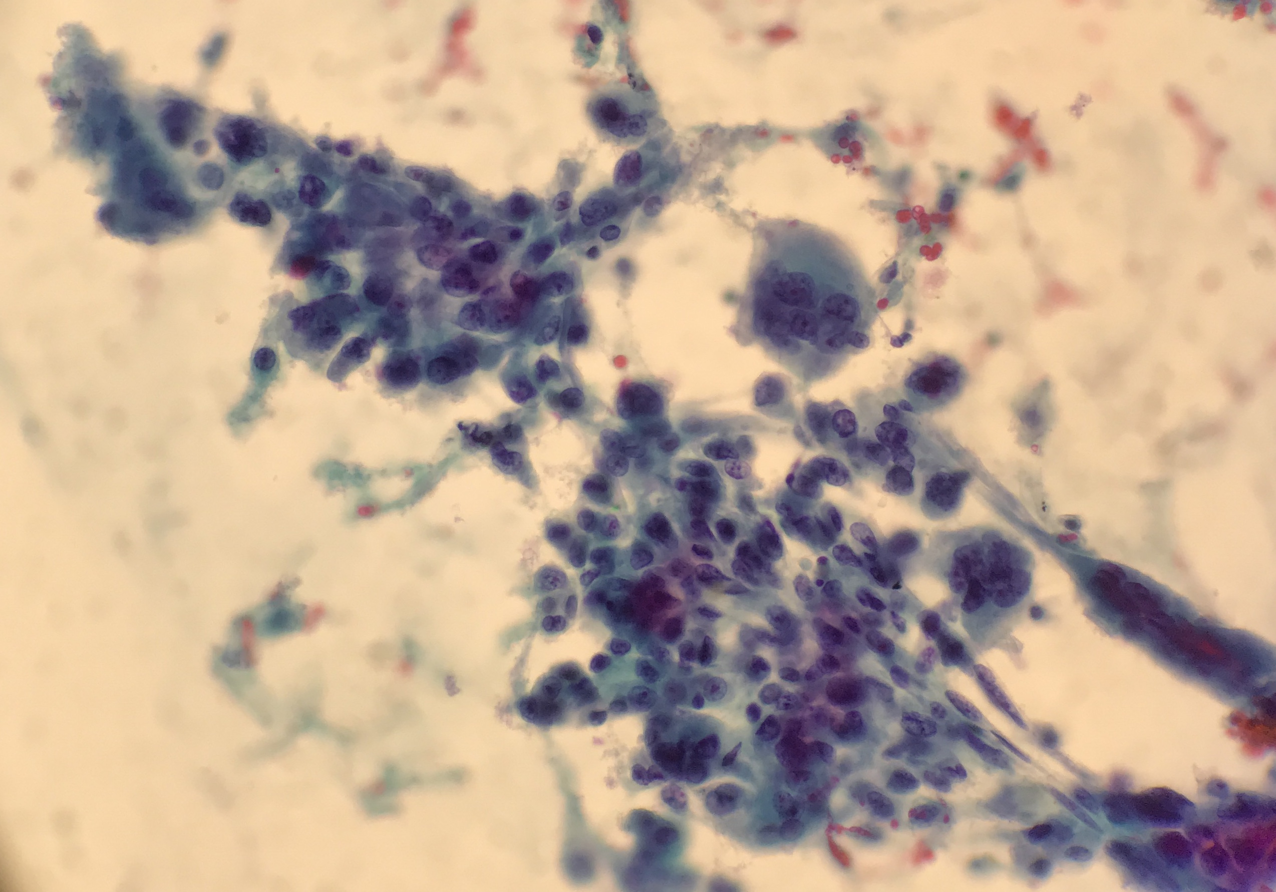
Figure 2: FNA Smear by Diff-Quik stain (400x)
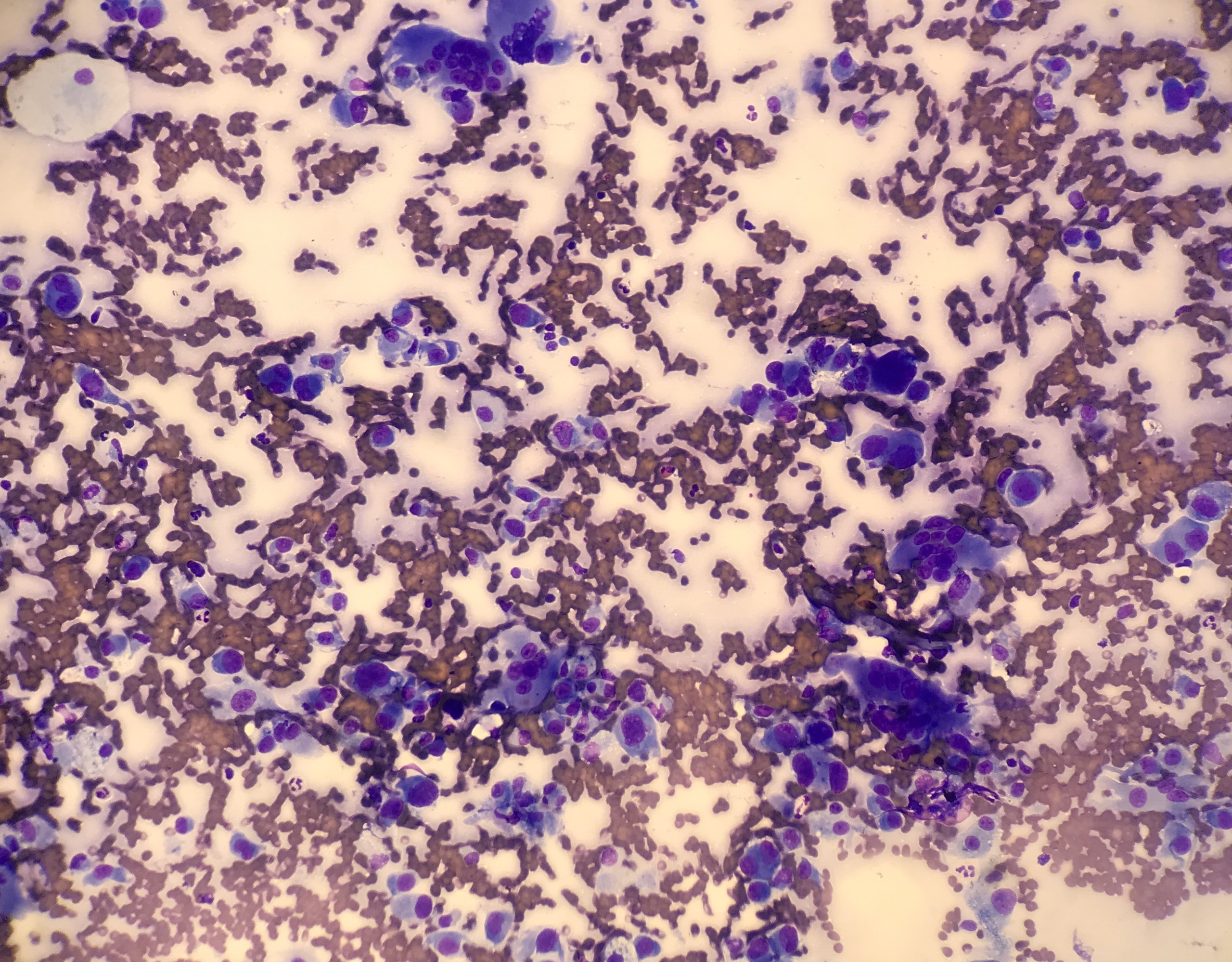
Figure 3: FNA Smear by Papanicolaou stain (200x)

Figure 4: FNA Smear by Papanicolaou stain (200x)
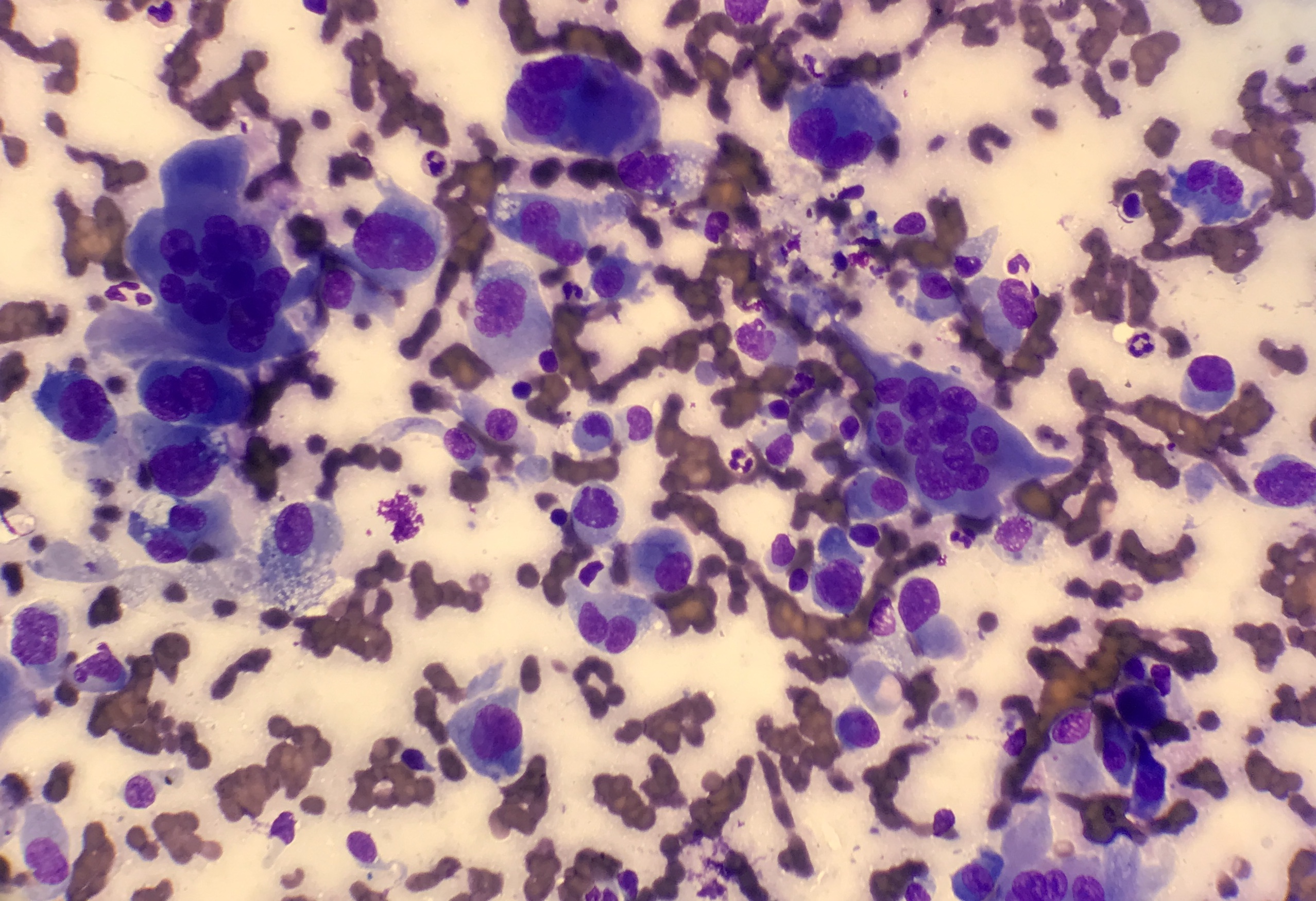
Figure 5: FNA Smear by Papanicolaou stain (400x)
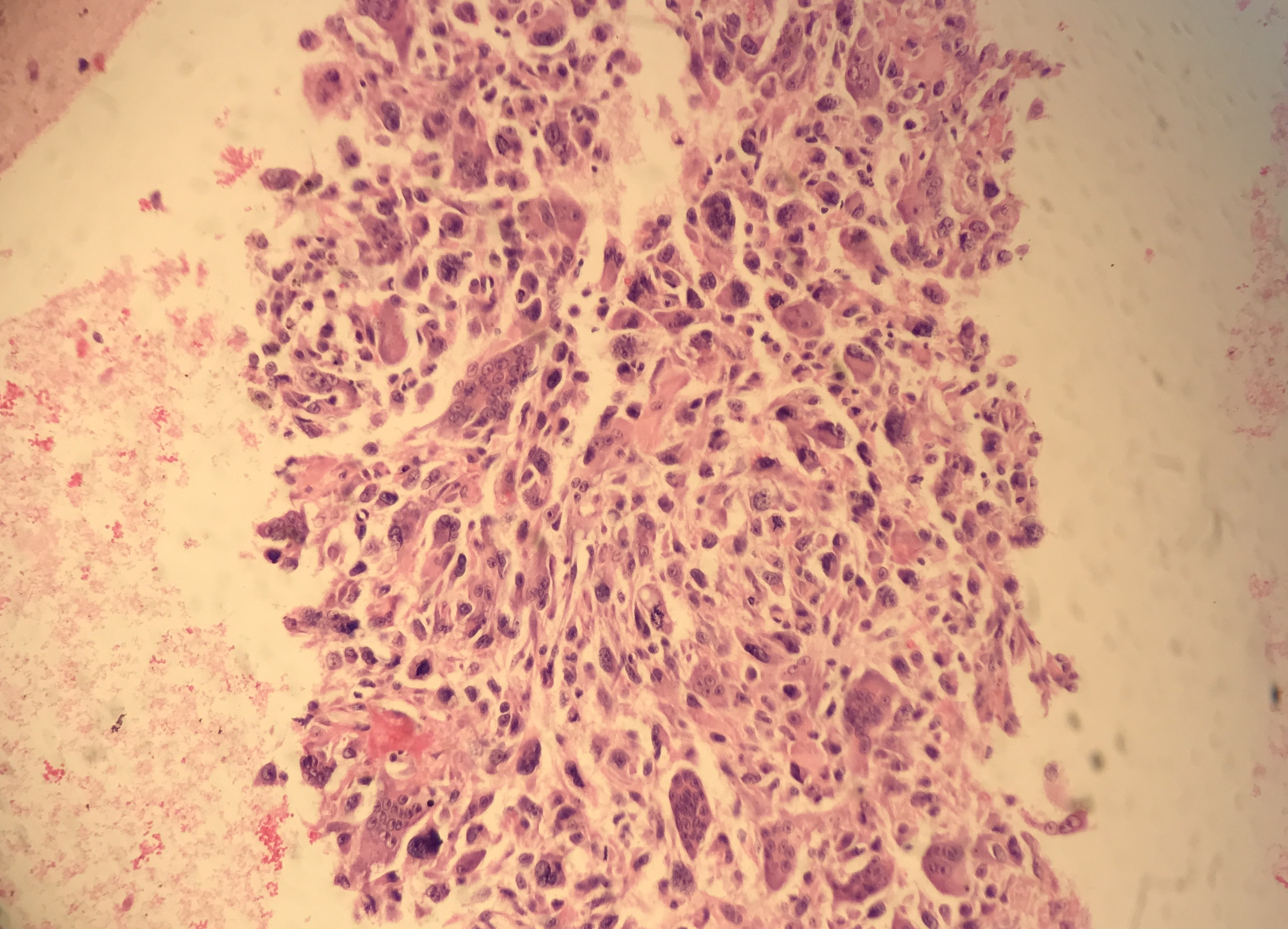
Figure 6: H&E staining on FNA cell block (200x)
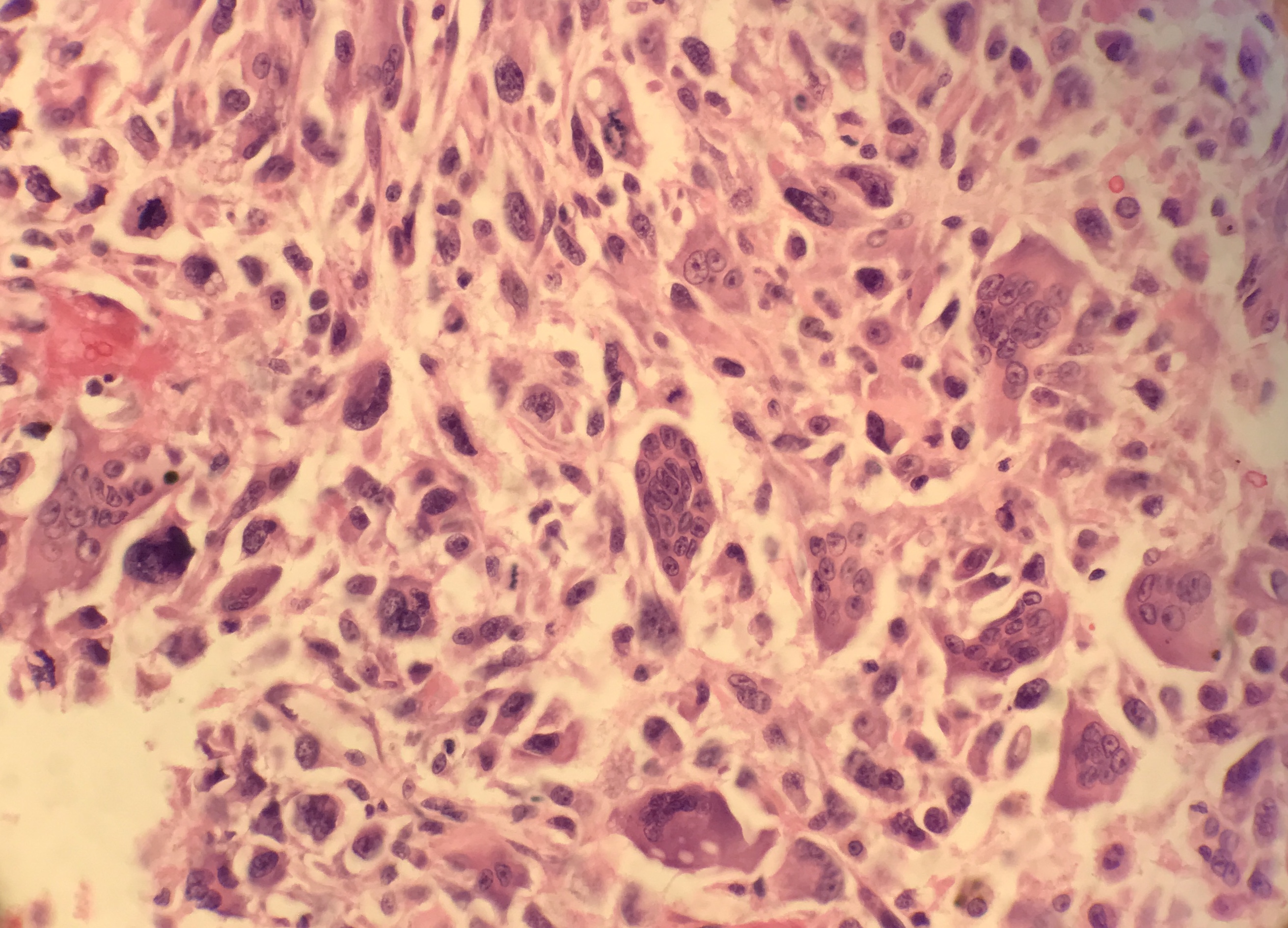
Figure 7: H&E staining on FNA cell block (400x)
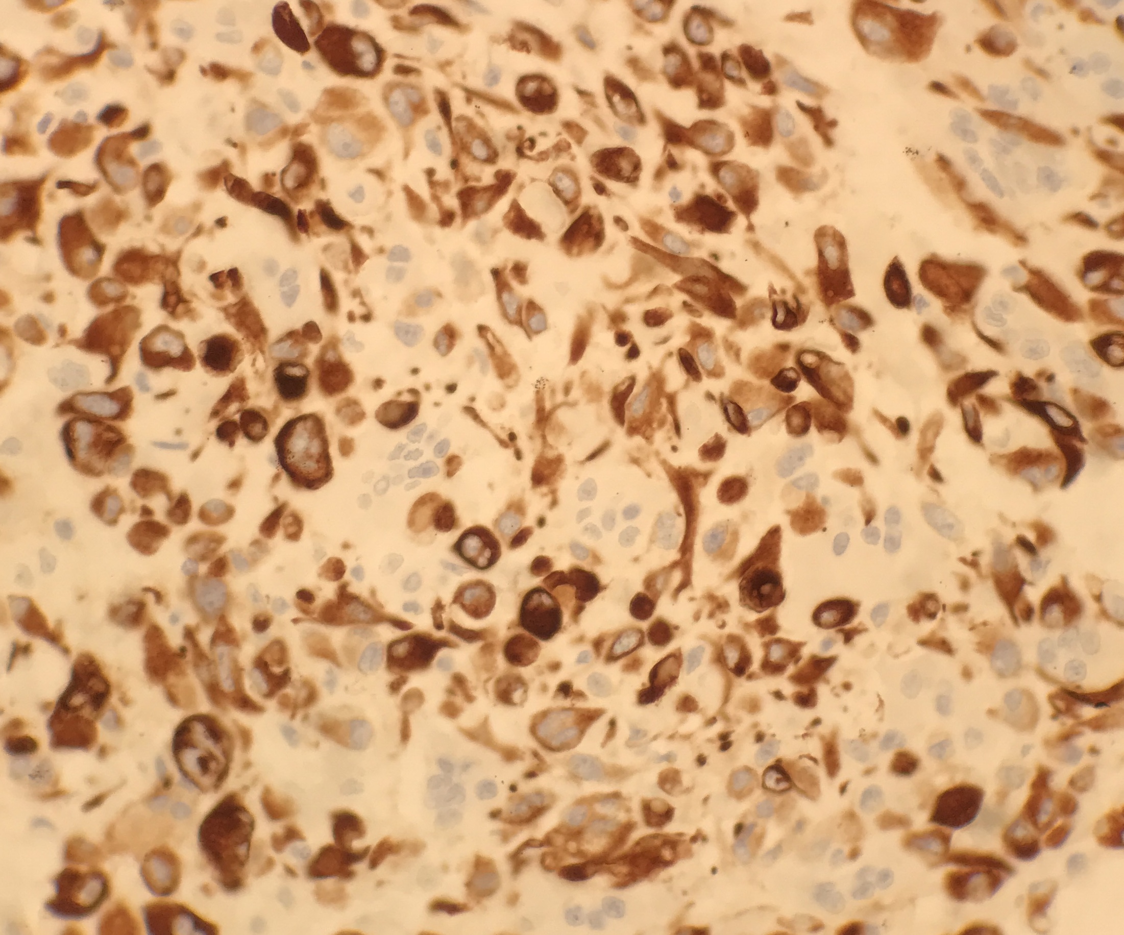
Figure 8: CK AE1-AE3 Immunohistological stain
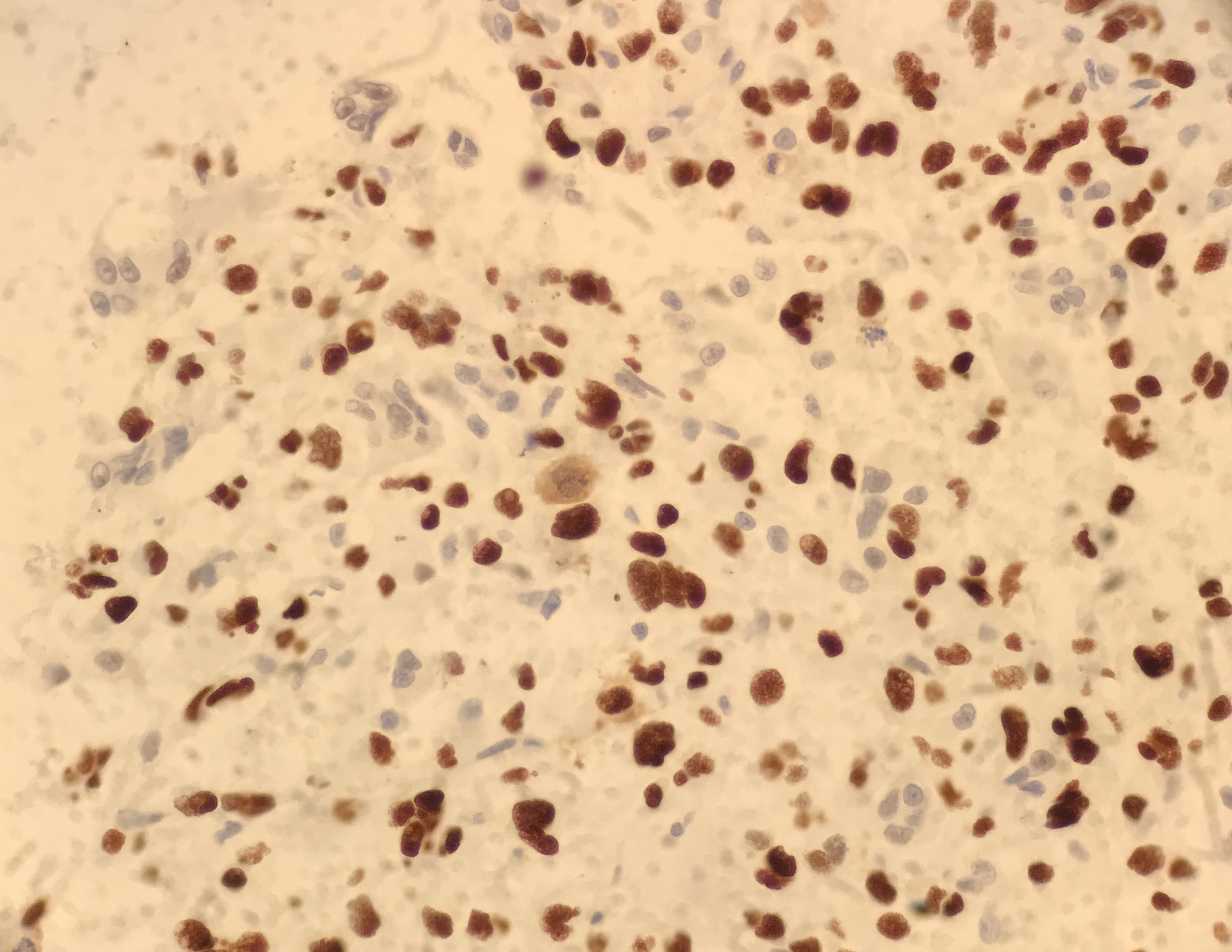
Figure 9: p53 Immunohistological stain on cell block
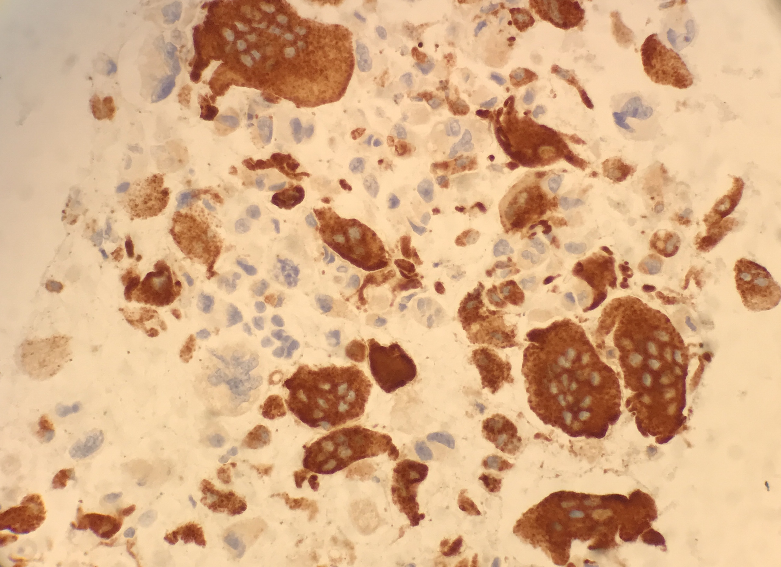
Figure 10: CD68 Immunohistological stain
The above features are compatible with which of the following?
Choose one answer and submit.
D.) Undifferentiated carcinoma of the pancreas with osteoclast-like giant cells
> Learn more about this diagnosis.
 Meet our Residency Program Director