
Residency Program - Case of the Month
July 2018 - Presented by Dongguang Wei (Mentored by Tao Wang)
Clinical History
A 17-year-old female presented with right femur fracture and underwent intermedullary nailing and open reduction internal fixation. She complained right hip pain again 9 months after the surgery. X-ray revealed hardwire failure and non-union of the fracture. Biopsy demonstrated only vascular tissue. She underwent revision with hardware replacement. Culture of the non-union fracture comes back negative. She complained of persistent right hip pain, X-ray showed right femoral neck fracture around the nail and she was admitted again for a right femur removal of hardware and proximal femoral replacement.
Further Imaging Studies
CT and X-ray shows intraosseous hemangioma, diffuse demineralization/osteolysis of the proximal femur, underlying severe osteoporosis and tapering of bone.
Pathology Review
Multiple pathology reviews showed fragments of viable bones with remodeling changes, negative for acute inflammation; Benign lymphovascular proliferation with network of thin-walled vessels highlighted by CD31, CD34, and D2-40. Osteoclasts are noted within the adjacent bones, and demonstrate scalloped osteoclastic activity.
Images
Click on images to enlarge.
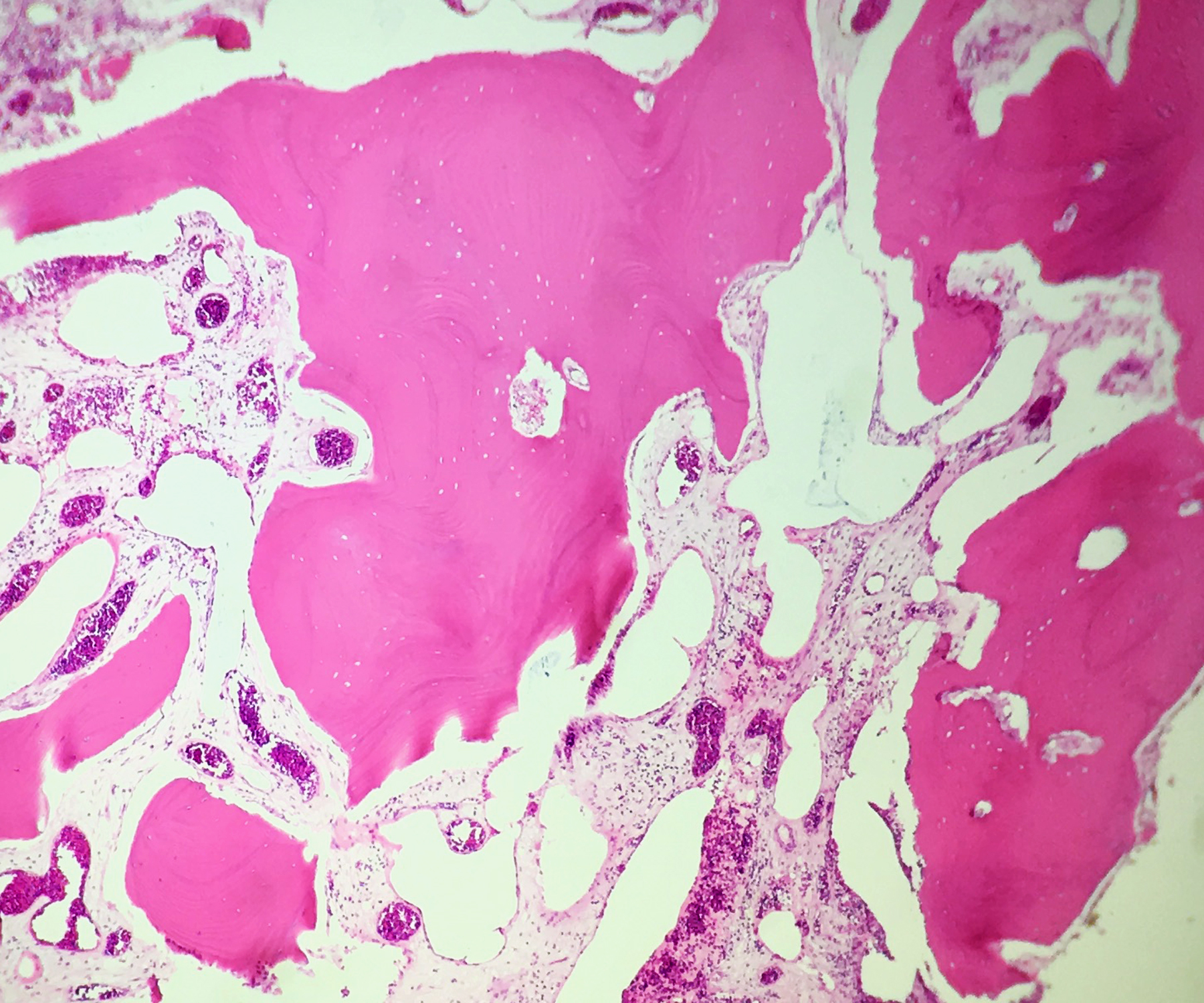
Figure 1: H&E 4x
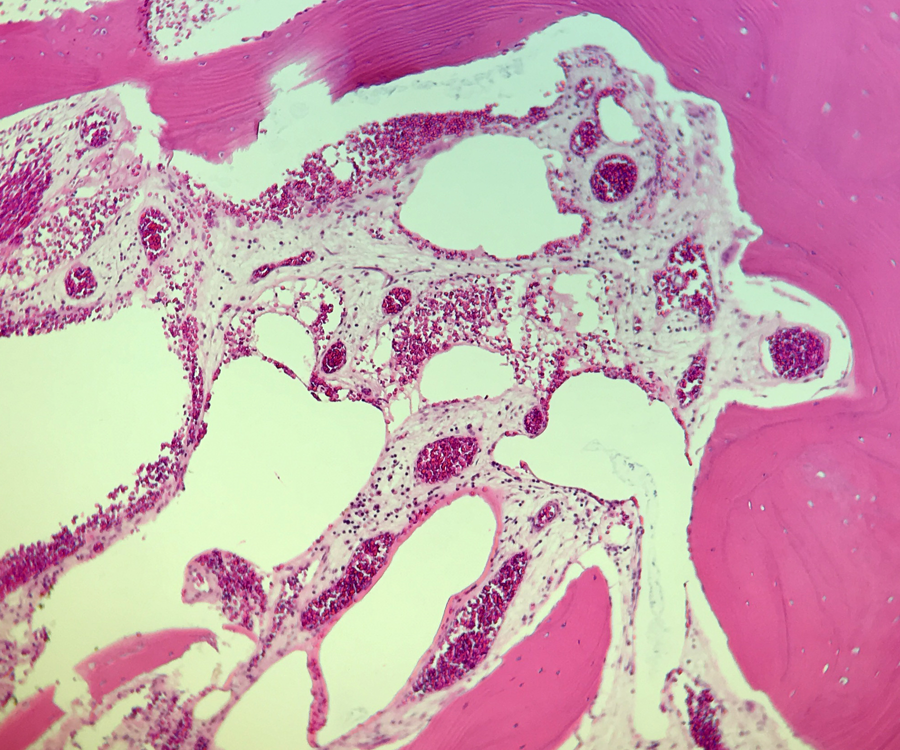
Figure 2: H&E 10x
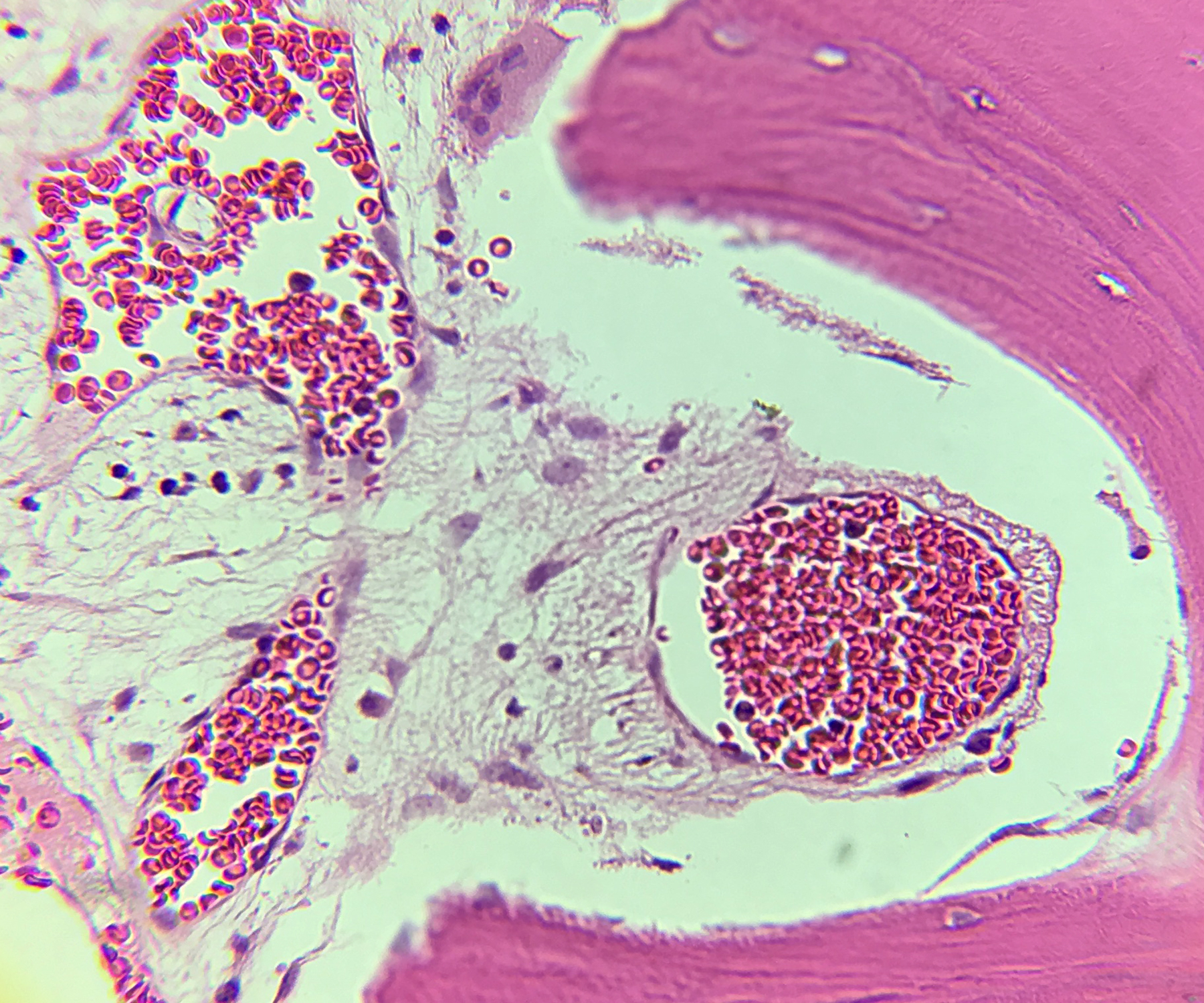
Figure 3: H&E 40x
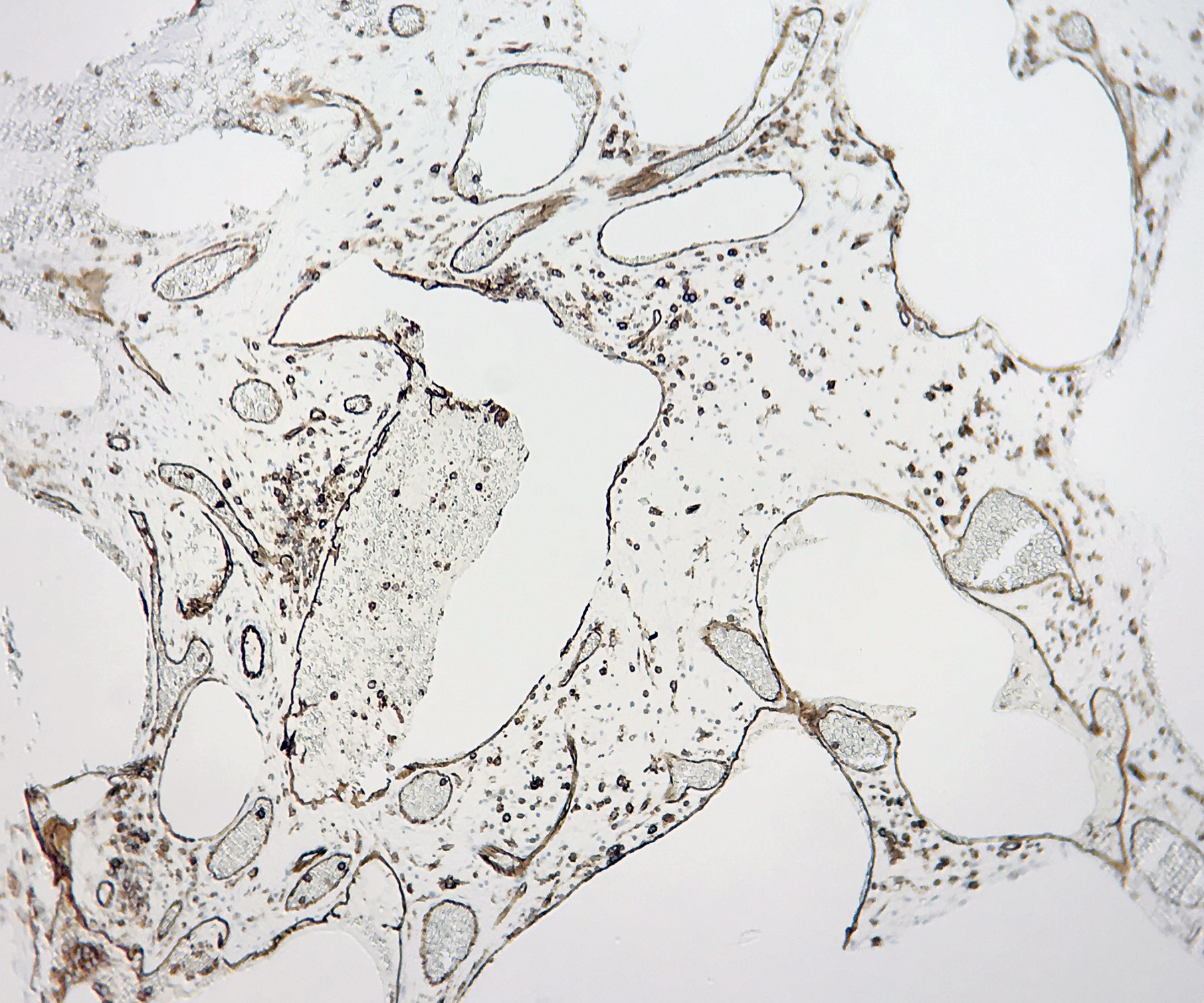
Figure 4: CD31 10x
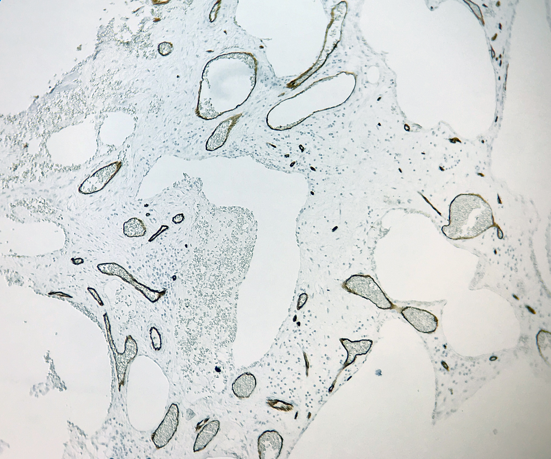
Figure 5: CD34, 10x
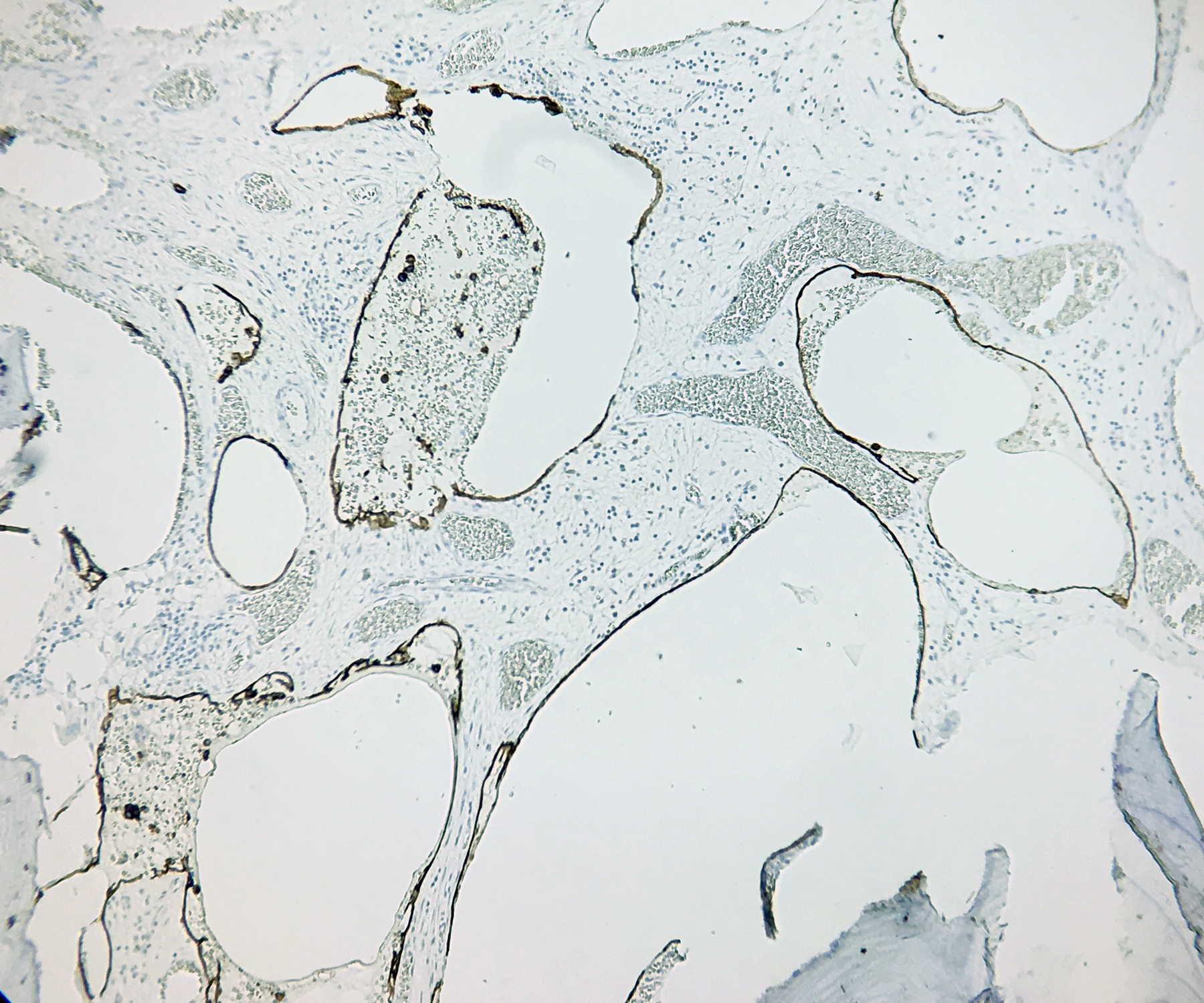
Figure 6: D2-40 10x
Which of the following is most likely the diagnosis?
Choose one answer and submit.
D. Gorham-Stout Syndrome (Vanishing Bone Disease)
> Learn more about this diagnosis.
 Meet our Residency Program Director