
Resident Program - Case of the Month
December 2018 - Presented by Tahera Iqbal (Mentored by Regina Gandour-Edwards)
Clinical History
A 66-year-old male presented at our institution for the evaluation of his right neck mass. After one month of the mass appearance, he started developing symptoms that included difficulty swallowing solids and change in voice. Ultrasound of his neck revealed an 18 mm slightly irregular hypoechoic mass in the submandibular gland. Adjacent to the mass was an elongated appearing lymph node with a fatty hilum measuring 2.7 X 0.9 X 0.7 cm. Patient reported of having thrush for four years, which made it hard for him to swallow. He admits to neck pain, but denies numbness or weakness of the face. Furthermore, he had no sinus symptoms, difficulty breathing, fever or unexpected weight loss. A biopsy specimen was taken from the mass and he subsequently underwent a submandibular gland excision with lymph node dissection.
Pathology Review
Gross examination revealed a 1.8 x 1.5 x 1.2 cm tan-gray mass within the salivary gland. The salivary gland weighed 9.1 g and measured 4.8 x 3 x 1.8 cm. The uninvolved cut surfaces are tan-brown and lobulated. Microscopically, the specimen showed markedly infiltrative islands of uniform cells of with mildly pleomorphic nuclei along with perineural invasion (Figure 1-3). 42 lymph nodes were examined, and they did not show tumor cells.
Click on image to enlarge.

Figure 1: H&E 4X
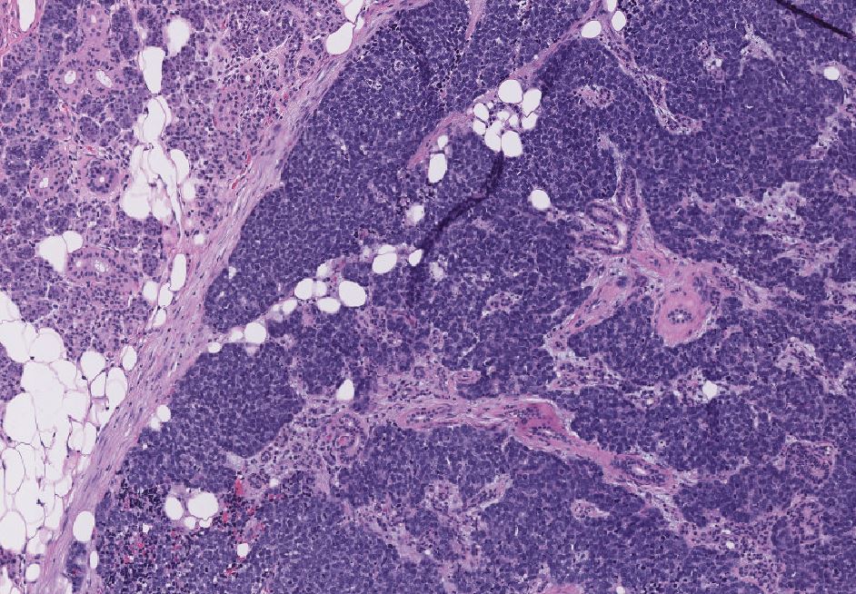
Figure 2: H&E 20X

Figure 3: H&E 20X
What is the diagnosis?
Choose one answer and submit.
A.) Adenoid Cystic Carcinoma with solid pattern predominance
> Learn more about this diagnosis.
 Meet our Residency Program Director