
June 2021 – Presented by Dr. Peter Michael Conner (Mentored by Dr. Regina Gandour-Edwards)
Clinical Findings
A 14-year-old female presented with a left neck swelling that has been present for several years. She initially thought it was normal and didn’t pursue care. A few months prior to presenting at UC Davis, she saw her PCP due to feeling tired and the noted the left parotid/left neck mass. Her past medical history includes a 7mm thyroid nodule.
Imaging Findings
An ultrasound showed a bilobed complex solid and cystic mass at the left submandibular region. A follow-up CT scan showed the mixed solid and cystic mass identified by ultrasound is located in the inferior portion of the left parotid gland. (Figure 1) The left submandibular and posterior cervical triangle lymph nodes on the left side are asymmetrically larger than their counterpart on the right however they remain within normal limits by CT size criteria and do not appear to be morphologically pathologic.
Macroscopic Findings
A left superficial parotidectomy was received which contained an ovoid, 3.6 x 1.5 x 1.3 cm pink and tan fleshy nodule.
Microscopic Findings
The tumor consists of medium to large, well encapsulated nodules containing large atypical cells with basophilic, granular cytoplasm and round, eccentric nuclei in glandular-like to microcystic growth patterns. Additionally, there is a prominent lymphoid inflammatory infiltrate. (Figures 2, 3, 4)
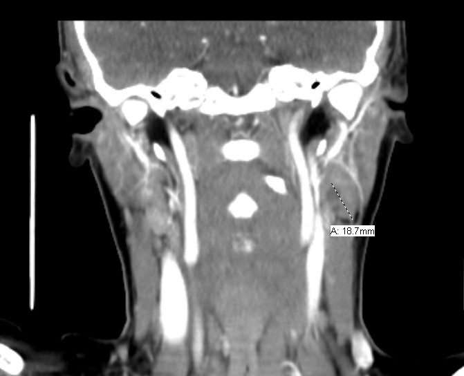
Figure 1: CT scan
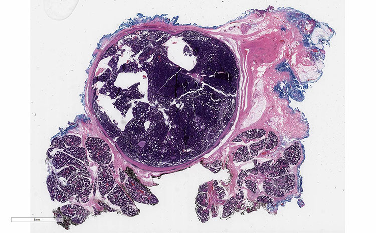
Figure 2. H&E low power 0.5x
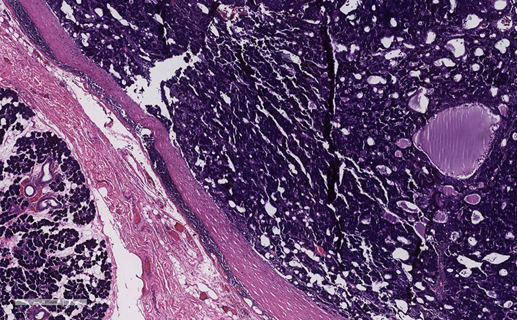
Figure 3. H&E Low power 4.0x
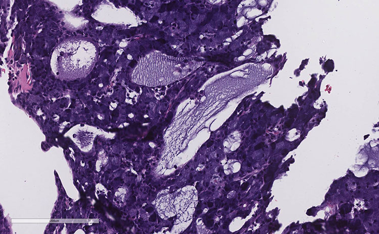
Figure 4. H&E High Power 20x
 Meet our Residency Program Director