
February 2022 – Presented by Dr. Sohrab C. Kadivar
A 48 year old male with no pertinent past medical history presented to the Emergency Department with progressive right flank and back pain, and gross hematuria. Review of systems was otherwise negative, including no unexplained weight loss. The patient reported a 12 pack year smoking history. Review of family history negative for urological cancer.
A CT scan of the abdomen and pelvis revealed a 7.8 by 7.6 by 7.3 centimeter heterogenous, hypoenhancing, mildly exophytic mass in the upper to mid pole of the right kidney. A central, low-attenuation focus was noted (measuring 4.0 by 3.0 centimeters) concerning for necrosis. The patient was scheduled for robot assisted laparoscopic radical right nephrectomy.
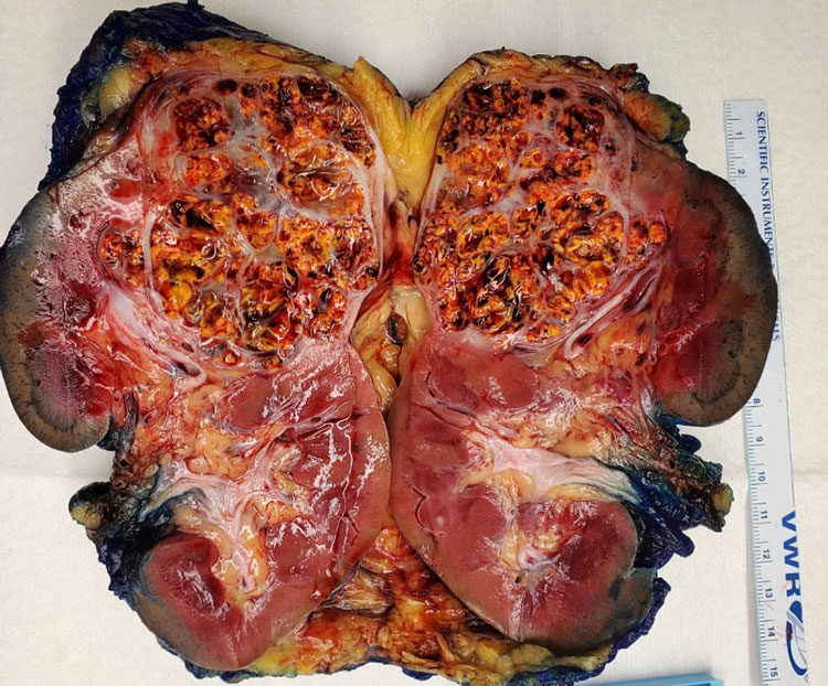
Gross examination of the kidney confirmed a single mass in the right upper pole measuring 6.1 by 5.9 by 4.3 centimeters. The tumor was haphazardly organized into numerous septations, interspersed with hemorrhage. There was no grossly identifiable extension of the tumor beyond the renal capsule.
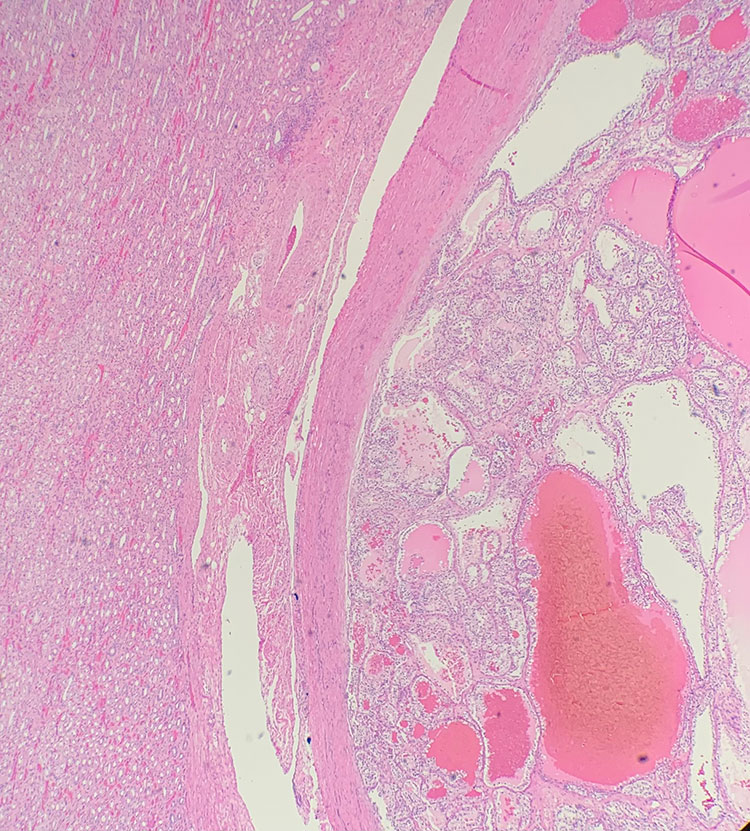
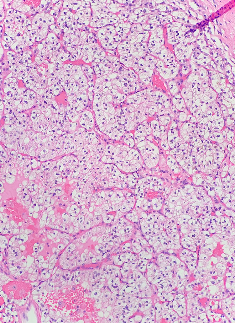
Histologic evaluation revealed grade two renal clear cell carcinoma, with no rhabdoid and no sarcomatoid features. Neither tumor necrosis nor lymphovascular invasion were identified. Microscopic examination confirmed the tumor was confined to the kidney, and surgical margins were negative for tumor.
> Learn more about this diagnosis.
 Meet our Residency Program Director