
Resident Program - Case of the Month
March 2022 – Presented by Jiejun Wu (Mentored by Han S. Lee)
Discussion
Myxopapillary ependymoma has some characteristic features to help its diagnosis and differentiation from classic type ependymoma: 1) Location, almost exclusively in region of conus medullaris, cauda equina and filum terminale as in this case (L1). 2) Share similar morphology with other ependymal tumors with tumor cells arranged around vascularized cores (perivascular pseudorosettes; Fig. 1 and 3). Differently, as the name indicated, myxopapillary ependymoma generally has myxoid stroma in these cores which has variable papillary architecture. 3) IHC profile show GFAP + for tumor cells (Fig. 2A), Alcian blue + for mucin, EMA ± with punctate (aka perinuclear dot-like, Figure 3B) reactivity. In this case, however, the EMA is negative (Fig. 2B). 4) In this case, mitotic index is low (Fig. 2C). However, following the 2021 CNS WHO classification, myxopapillary ependymoma is now regarded as a grade 2 neoplasm (Ref. 1). Anaplastic examples are exceptional.
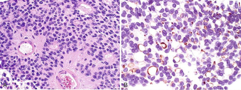
Figure 3. Ependymoma, classic type, WHO Grade II. A, H&E. B, EMA with EMA-positive dot-like structures (From Ref. 2)
Subependymoma is a low-grade (WHO Grade I) glioma often found intraventricularlly. One of its morphological features is small clusters of bland glia cells embedded in abundant fibrillary matrix with frequent microcystic change and sometimes calcification. GFAP is positive but EMA is negative (unlike ependymoma or myxopapillary ependymomoa).
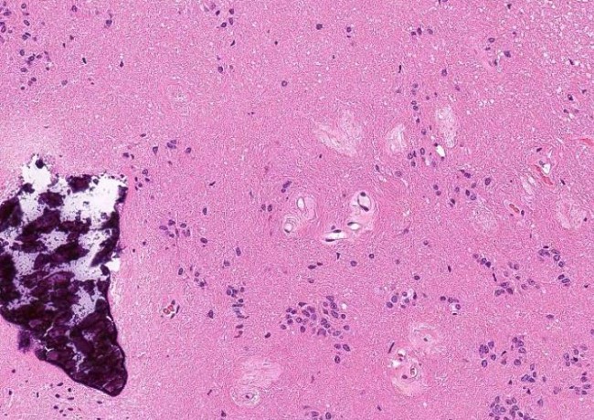
Figure 4. Subependymoma. Bland glia cells with abundant fibrillary matrix, frequent microcysts and sometimes calcification (from Ref. 3)
At this location, other differential entities should include schwannoma and paraganglioma. Schwannoma has its typical features of Antoni A and B tissue, Verocay bodies, diffuse and strong S100 and focal or negative GFAP. Paraganglioma has its typical neuroendocrine morphology without myxoid background but with positive neuronal markers.
References
- "WHO Classification of Tumours Editorial Board". World Health Organization Classification of Tumours of the Central Nervous System. 5th ed. Lyon: International Agency for Research on Cancer; 2021.
- Martin SE et al. "Neuropathology". In: Chen L, Bostwick DG, editors. Essentials of Anatomic Pathology, 4th, Springer International Publishing, Cham, Switzerland, 2016. p. 667-749.
- "Schaberg K. Kurt’s notes.", accessed on 02/17/2022
 Meet our Residency Program Director