Resident Program - Case of the Month
November 2022 – Presented by Dr. Jiejun Wu (Mentored by Dr. Kurt Schaberg)
Discussion
The correct answer is B. The diagnosis is a littoral cell angioma. In this case, representative sections from splenectomy show a benign vascular proliferation comprised of anastomosing vascular spaces, some with cyst-like spaces with irregular lumina. The lining cells are composed of tall endothelial cells. Many vascular lumina appear to contain sloughed endothelial cells and cellular debris. No atypia is noted. No necrosis is present. No mitoses are seen. In addition, there are many scattered megakaryocytes and possibly rare scattered erythroid precursors.
Littoral cell angioma is an extremely rare benign splenic tumor with typical histomorphologic and immunohistochemistry features. The lesional cells originate from littoral cells, the endothelial lining cells of red-pulp sinuses. Littoral cell angiomas, as shown in this case, present with unique splenic vascular neoplasms composed of anastomosing vascular channels and often have papillary projections and cyst-like spaces (Fig 1A). The endothelial cells frequently detach into the vascular spaces (Fig. 1B) and show evidence of hemophagocytosis (Fig. 1C) and extramedullary hematopoiesis (Fig. 1B).
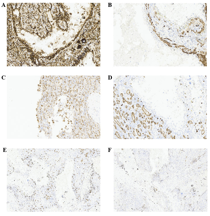
Immunohistochemistry staining is very helpful to the diagnosis of littoral cell angiomas. Unlike lesions from splenic cord capillary, littoral cell angiomas express endothelial marker CD31 but not CD34 (Fig 2A and 2B; Answer A and B). They also express histiocytic markers, such as CD68, CD163 and lysozyme (Fig. 2C; Answer C). Though CD8 staining is positive for sinus endothelial cells in normal spleen, the lesional cells in littoral cell angioma are often negative for this marker (Fig. 2D; Answer D). If, extramedullary is present, markers for megakaryocytes and erythroid precursors can also be positive (Figure 2E and 2F; Answer E and F). Pan cytokeratin maker such as AE1/AE3 is negative (Answer G).
 Figure 2. Immunohistochemistry staining.
A, CD31, diffusely positive. 200x. B, CD34, negative for the lining lesional cells. 200x. C, CD68, positive. 200x. D, CD8, negative for the lining lesional cells. 200x. E, CD61. 100x. F, E-cadherin. 100x.
This tumor is only found in spleen and is benign. There is no documented evidence of typical littoral cell angioma prior to malignancy development although there are cases of littoral cell angiosarcoma and littoral cell hemangioendothelioma which is designated as a transitional entity between these two. Other differential diagnoses include benign hemangioma, lymphangioma and malignant angiosarcoma, hemangioendothelioma and Kaposi sarcoma. Non-neoplastic malformations, reactive changes and infections should also be ruled out.
References
- Falk S, et al. Littoral cell angioma: a novel splenic vascular lesion demonstrating histiocytic differentiation. Am J Surg Pathol. 1991 Nov;15(11):1023-33.
- Aguilera N. Littoral Cell Angioma. accessed on 10/22/2022.
- Burke JS. Spleen. In: Longacre TA, editors. Mills and Sternberg's Diagnostic Surgical Pathology, 7th, Wolters Kluwer, 2022. p. 884

 Meet our Residency Program Director
