
December 2022 – Presented by Dr. Luke Dang (Mentored by Drs. Kurt Schaberg and Han Lee)
Clinical History
A 19 year-old female presents with a multiple year history of a palpable mass in the sacrococcygeal region overlying the coccyx and extending bilaterally to the intergluteal cleft, with recent interval growth.
Gross Examination
After resection, the received specimen weighs 760 grams and is comprised of skin and underlying soft tissue, including a mass which measures 13.1 x 11.1 x 8.2 cm. The mass abuts the superficial skin and deep margins, is lobulated in architecture with well-circumscribed borders, and has pink-tan, semi-firm, focally hemorrhagic cut surfaces (Figure 1).
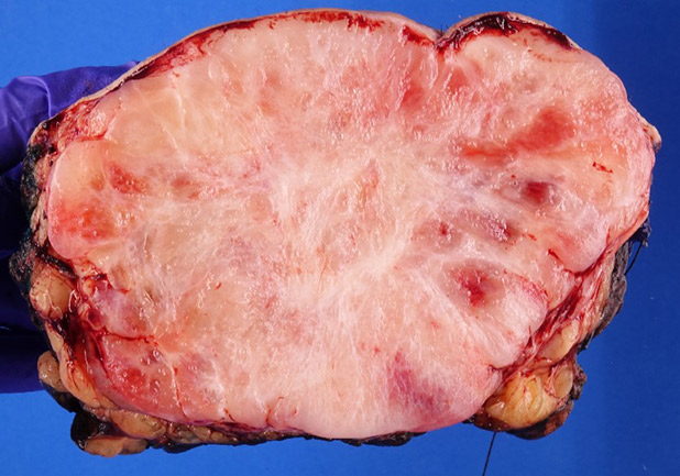 Figure 1. Representative Cut Surface of Mass
Microscopic Features
Histologic sections of the mass demonstrate prominent background myxoid matrix containing variable amounts of well-differentiated epithelial cells with eosinophilic cytoplasm and bland oval nuclei (Figures 2-4). In some areas, these epithelial cells line hyalinized vascular channels and surround microcysts containing mucin (Figures 3-4).
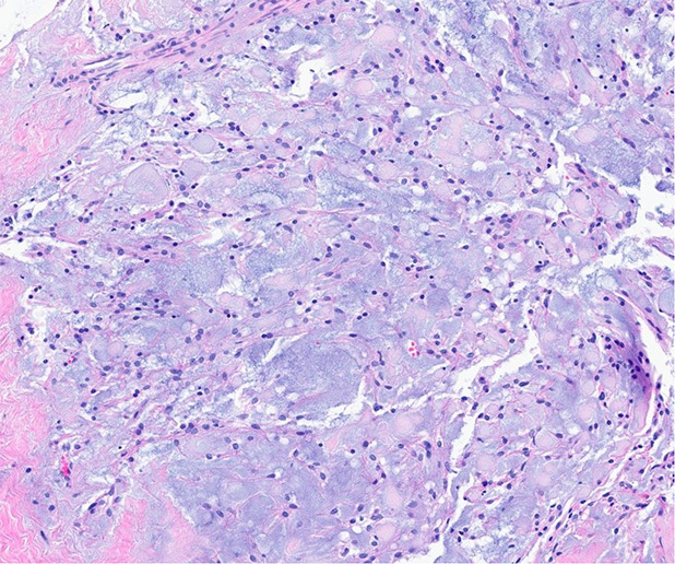 Figure 2. H&E, 10x.
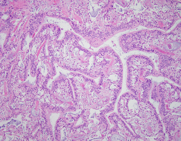 Figure 3. H&E, 10x.s
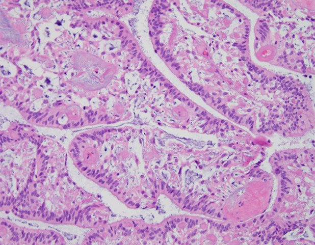 Figure 4. H&E, 20x.
Immunohistochemistry
Immunohistochemical (IHC) staining demonstrates that the specimen is positive for GFAP (Figure 4), variably positive for S100 (Figure 5), and negative for AE1/AE3 (pancytokeratin), EMA, ERG, CK7, SOX-10, brachyury and CK20. A low mitotic rate is demonstrated by Ki-67 IHC.
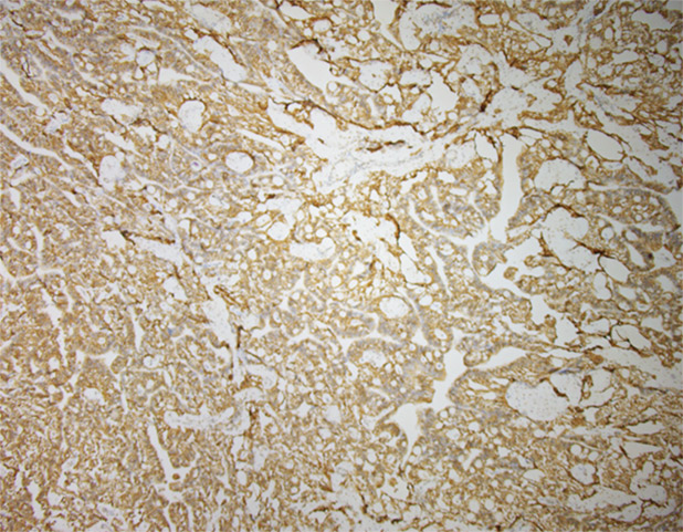 Figure 5. GFAP Immunohistochemistry.
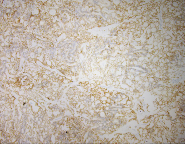 Figure 6. S100 Immunohistochemistry.
 Meet our Residency Program Director