
Resident Program - Case of the Month
February 2023 – Presented by Dr. Ahresh Saha (Mentored by Dr. Frank Melgoza)
Discussion
Histologic evaluation revealed grade 3 clear cell renal cell carcinoma. There were no rhabdoid or sarcomatoid features. Tumor necrosis and lymphovascular invasion were not identified. The capsular, peri-nephric fat, and parenchymal margins were clear of tumor. Osseous metasplasia was also identified arising in the clear cell renal cell carcinoma.
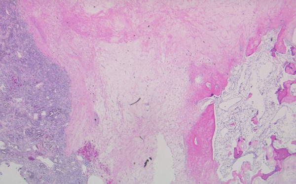 Picture of tumor (left side) next to osseous metaplasia
In tumors of the kidney, clear cell renal cell carcinoma is the most common occuring type. Histologically, the tumor is composed of compact nests and sheets of cells with clear cytoplasm and distinct membranes. The WHO grading system has 4 tiers and uses nucleolar prominence for its grading2.
Osseous metaplasia is the presence of bone formation. Osseous metaplasia with clear cell renal cell carcinoma (RCC) is exceedingly rare. There are less than 20 reported cases of osseous metaplasia in association with RCC1. The exact mechanism of bone formation in RCC is not known. Several hypotheses have been put forward for ossification in RCC. One hypothesis suggests simple production of bone by tumor cells secondary to ischemia, necrosis, inflammation or there might be ossification in preexisting focus of calcification1.
References
- Agarwal S, Bohara S, Jha R, Khurana N, Agarwal PN. Clear cell renal cell carcinoma with osseous metaplasia: Rare case report. J Cancer Res Ther. 2015 Oct-Dec;11(4):1039. doi: 10.4103/0973-1482.146109. PMID: 26881653.
- Nezami BG, MacLennan G. Clear cell. PathologyOutlines.com website. Accessed January 25th, 2023.
 Meet our Residency Program Director