
March 2023 – Presented by Dr. Yuden Pemba (Mentored by Dr. Maija Kiuru)
Clinical History
A 91-year-old female presented with a 3-month history of an itchy rash on her left thigh which was initially thought to be scabies. She was treated with oral ivermectin as well as permethrin cream. The rash improved initially, but then she started to notice blisters slowly appearing on other areas. She was referred to a dermatology evaluation.
Physical examination revealed urticarial plaques with some small intact blisters and excoriations on the bilateral thighs, buttocks, forearms, upper chest, and lower legs. Limited erosions were noted on the right buccal mucosa. There was no ocular involvement.
Microscopic Findings
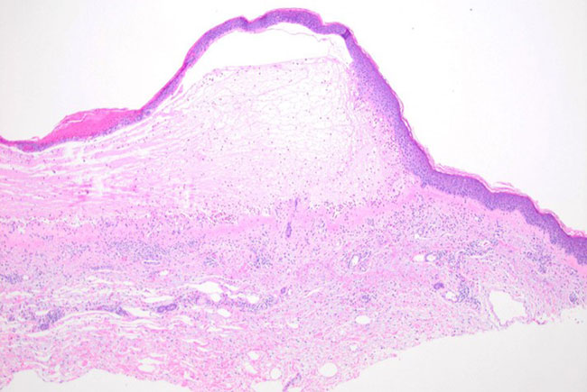 Figure 1. Subepidermal split between the epidermis and the dermis. (4x)
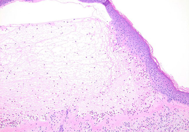 Figure 2. Close up of the subepidermal bulla with prominent eosinophils. (10x)
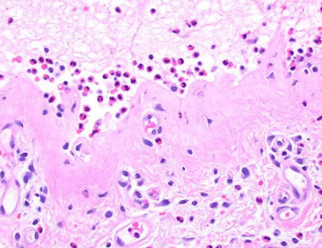 Figure 3. Inflammatory cells of eosinophils, neutrophils, and lymphocytes within the blister cavity and in the dermis (40x)
Direct immunofluorescence test showed a linear deposition of IgG and C3 along the basement membrane zone.
 Meet our Residency Program Director