Residency Program - Case of the Month
January 2010 - Presented by Diane Sanders, M.D.
Clinical history:
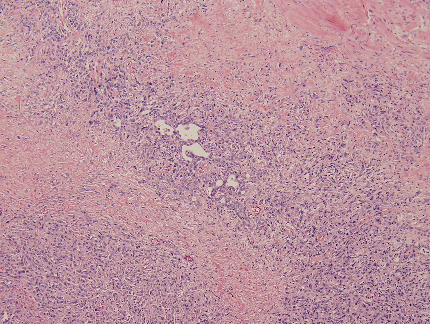
The patient is an 80 year-old woman who presented with a three month history of multiple urinary tract infections and one day of gross hematuria. She had a 15 pack year history of smoking. A CT scan revealed a bladder wall tumor with no evidence of metastases. Cystoscopy demonstrated a golf ball sized, exophytic tumor embedded in the right dome and wall of the bladder. After evaluation of biopsies and a TURBT, a radical cystectomy with bilateral pelvic lymph node dissection was performed.
Gross description:
A 3.0 x 3.0 cm partially ulcerated and fungating tumor was identified covering the dome and posterior wall of the bladder. The tumor was 1.3 cm thick and grossly extended through the muscular wall, abutting overlying adipose tissue.
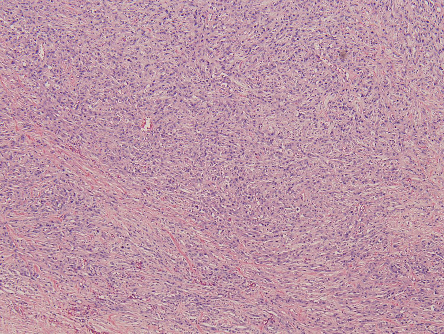
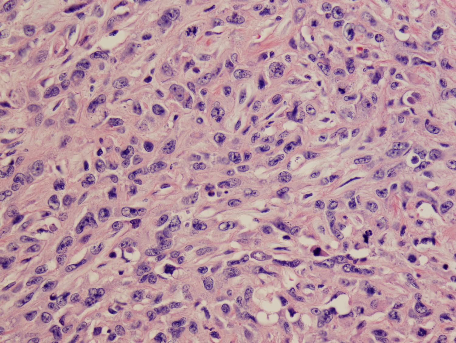
Microscopic photographs:
Immunohistochemical Stains:
| Vimentin: | Positive in sarcomatous areas |
| p63: | Weakly positive |
| CK20: | Negative |
| CK7: | Positive in epithelial areas |
| Villin: | Negative |
|
Figure 1: Vimentin  |
Figure 2: CK7 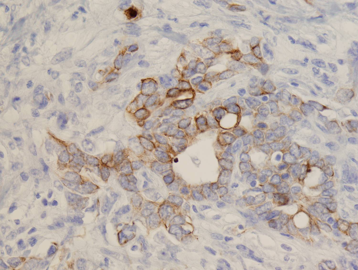 |
Figure 3: p63 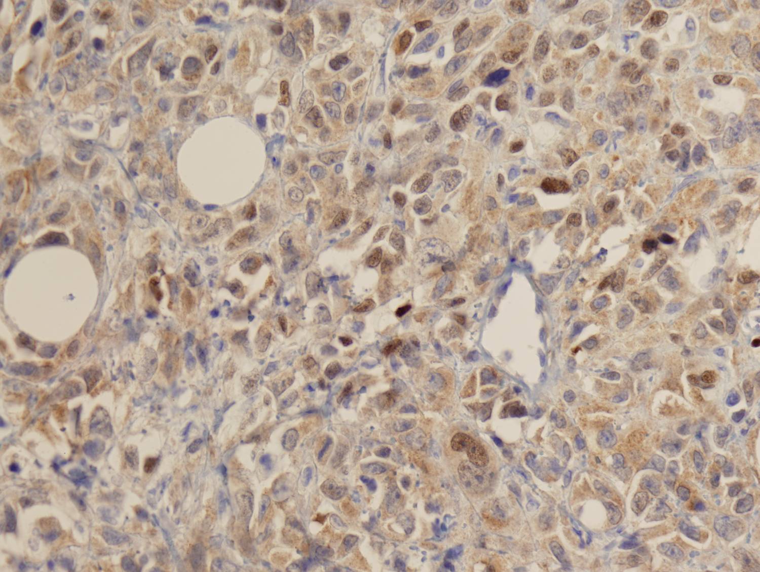 |

 Meet our Residency Program Director
