Residency Program - Case of the Month
July 2010 - Presented by Tatyana Berezenko, M.D.
Clinical history:
Patient is a 34 year old male with no significant past medical history who presented with right painful lump, behind right ear for 2.5 months. The ultrasound of the right tale of his parotid gland showed a 3.0 x 2.6 x 1.6 cm irregular hypoechoic mass. The fine needle aspiration revealed cytological atypia. A CT ordered for further evaluation showed a fairly well-circumscribed mass in the superficial portion of right parotid gland, measures 3.0 cm in greatest dimension. The patient underwent right parotidectomy.
Gross description:
The right parotidectomy specimen revealed a well circumscribed solid cystic mass with cyst, measures 1.9 x 1.6 x 1.7 cm, containing bloody fluid. The mass has cut surfaces that are tan-red in color with foci of hemorrhage noted and cyst formation ranging in size from 0.2 cm to as large as 0.9 cm.
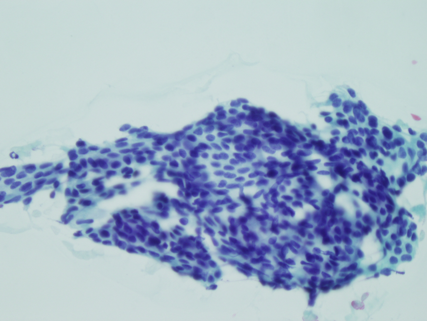
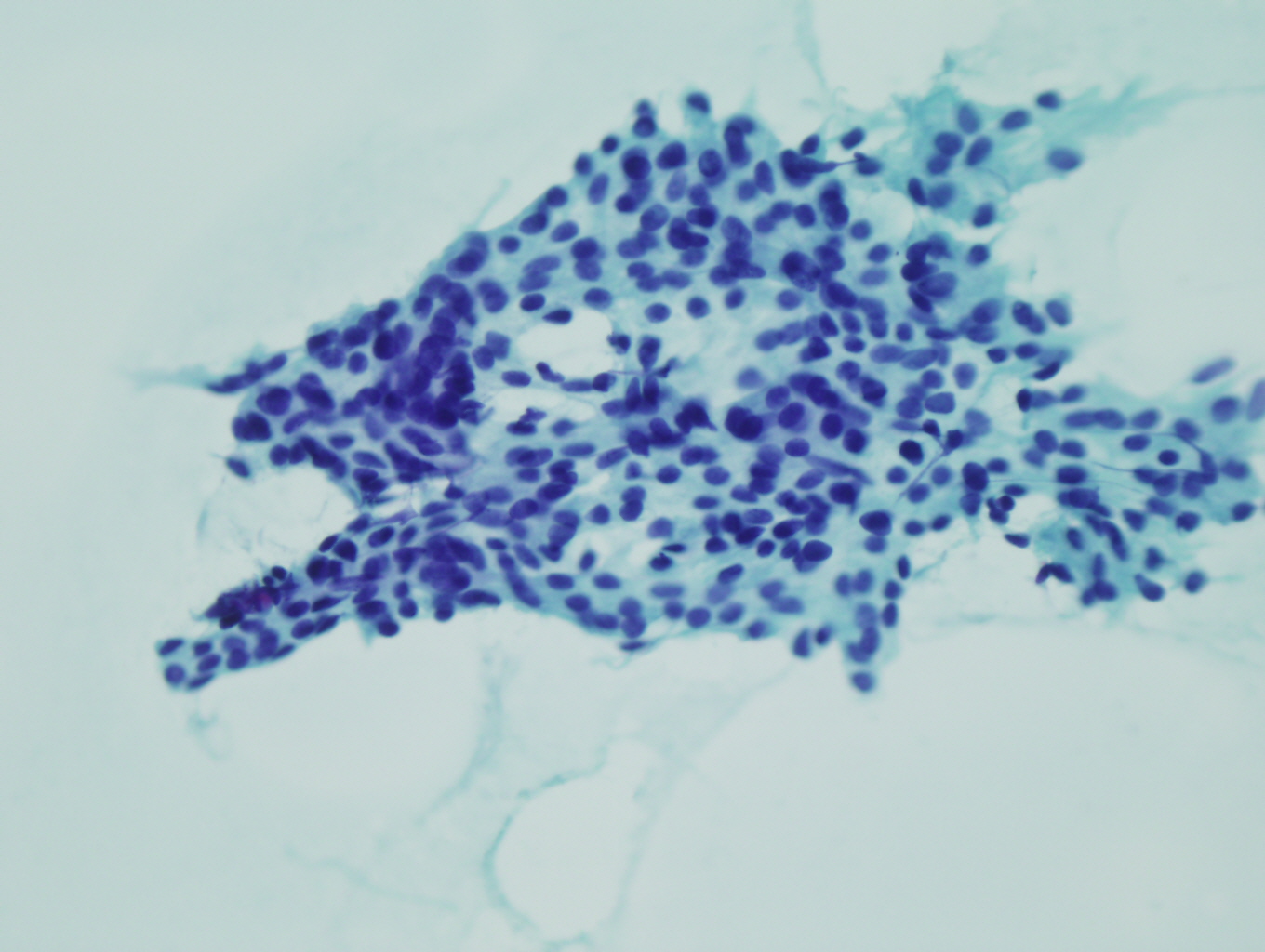
FNA:
Microscopic Photographs:
Microscopically the tumor showed varying histologic patterns (figures 1-6) with the predominant pattern shown in figures 3-5.


 Meet our Residency Program Director
