Residency Program - Case of the Month
December 2011 - Presented by Meighan Tomic, M.D.
Clinical history:
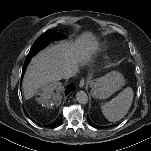
The patient is a 64 year old female, with a past medical history significant for chronic obstructive pulmonary disease, diabetes mellitus, and hypertension, who was found to have a right adrenal mass on chest CT (during work-up for pneumonia). Follow up abdominal CT showed a well circumscribed, 6.6 x 7.2 x 7.5 cm, mass with areas of high tissue density, fat density and multiple dystrophic calcifications. She subsequently underwent a right adrenalectomy.
Abdominal MRI:
Gross description:
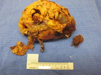
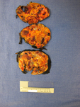
Received fresh was a 152 gram adrenal gland (10.3 x 6.8 x 5.2 cm) with scant attached fat. The yellow-orange surface was partially disrupted. Cut surfaces varied in appearance with solid, golden-orange areas and cystic areas (0.3 - 2.0 cm). The cysts were filled with gelatinous gray to hemorrhagic material. No definitive residual normal adrenal gland was identified.
Gross images:
 |
 |
Microscopic photographs:
 |
 |

 Meet our Residency Program Director
