Residency Program - Case of the Month
February 2012 - Presented by Kali Tu, M.D.
Clinical history:
A sixteen year old girl with no significant past medical history presented with a tender right breast lump. She was initially treated with antibiotics; however the mass persisted with increased pain and tenderness. A mammogram detected an infiltrative and hypoechoic 5.2cm mass at the 10 o’clock position. Complete history revealed two episodes of epistaxis and one episode of blood in her stool in the past week and physical exam revealed petechia on her lower legs. Complete blood count showed pancytopenia. Additional imaging showed a sub-acute left foot fracture.
| WBC: | 4.4 k/mm3 (4.5-13.5k/mm), 3% blasts (by flow cytometry) | |
| Hemoglobin: | 9.0 gm/dL (11.5-15.5gm/dL) | |
| Hematocrit: | 25% (35-45%) | |
| Platelet: | 34 k/mm3 (130-400 k/mm3) |
Histologic descriptions:
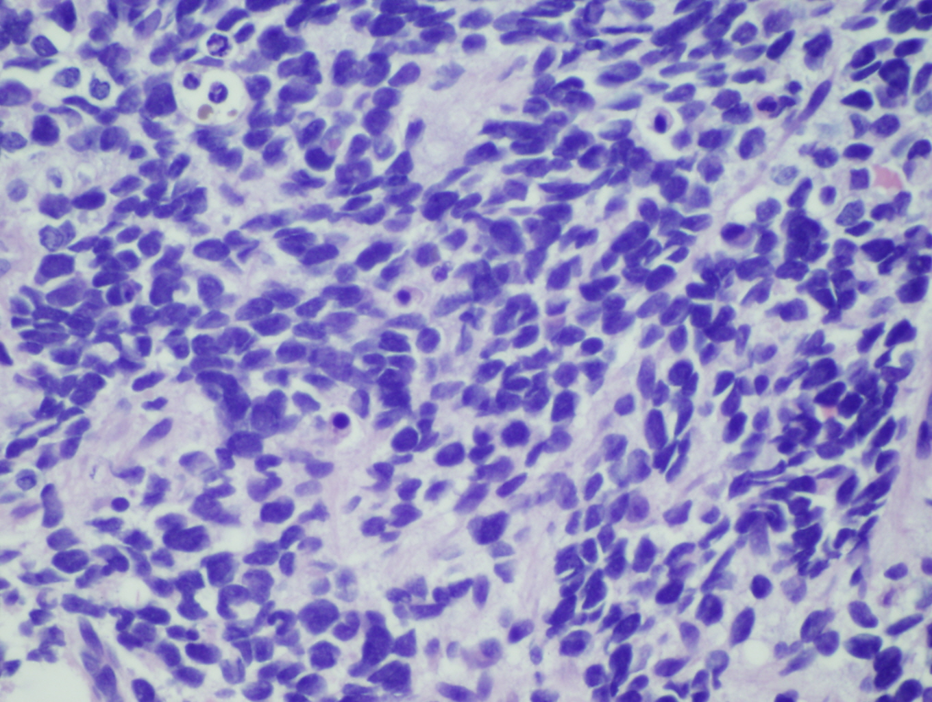
- Breast biopsy: H&E 2x, 60x. The low-power image shows a small amount of residual adipose tissue and fibrous stroma. Most of the tissue has been infiltrated by sheets of small round blue cells. On high-power these cells are easily identified as monomorphic small cells in sheets and cords with high nuclear to cytoplasmic ratio. Some cells show abundant eosinophilic cytoplasm. The nuclei are pleomorphic and some have a prominent nucleolus. Mitosis and pyknotic cells are easily identified.
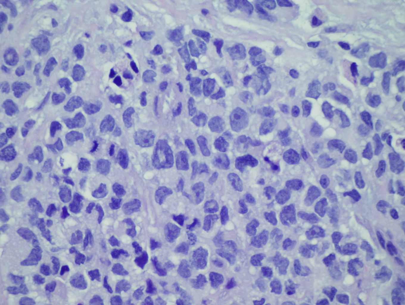
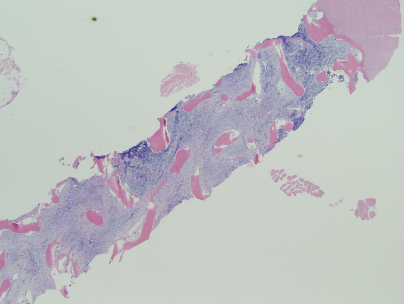
- Bone marrow: H&E 2x & 60x. The low-power images show the bone marrow is completely replaced by sheets of cells with an eosinophilic stroma. On high-power, there are small round blue cells in sheets, nests and cords. Some of the cells have an eccentrically located nucleus with a small amount of eosinophilic cytoplasm. Nucleoli are inconspicuous and mitotic figures are easily identified.
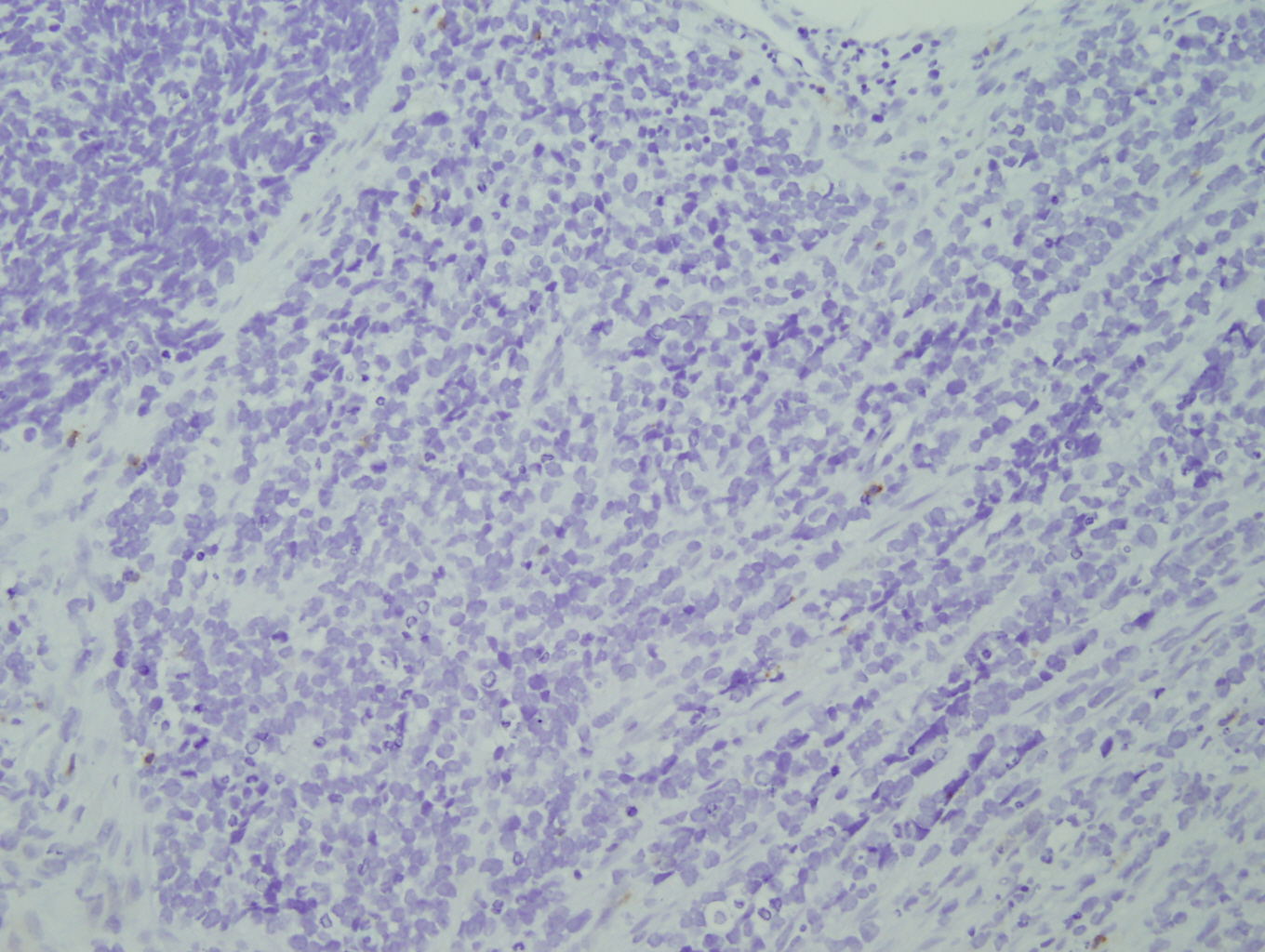
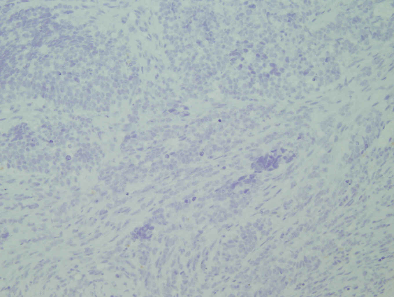
- AE1/AE3: Negative.
- CD45: Negative.
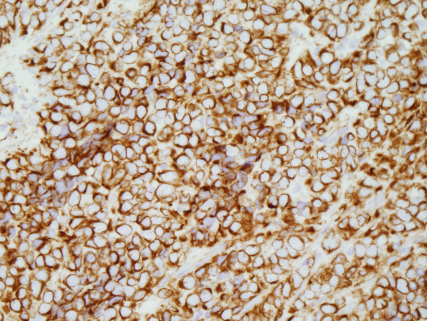
- CD56: Positive
- CD57: The cells show focal positivity.
- Desmin/Myo-D: Positive.
- Ki-67: The cells show a high proliferative index about 70%.
- WT-1: The cells show uniform and strong cytoplasmic staining
- Additional negative stains: EBER.
Flow cytometry:
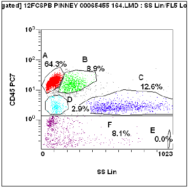
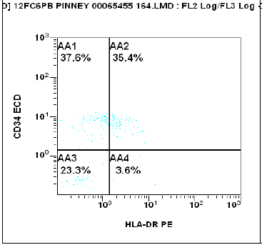
In the CD45 dim gate (gate “D” blast gate) there is a distinct population of cells expressing CD34+/ HLA-DR+/CD33+ dim. The cells were also CD117+ (not illustrated here). This is the phenotype of myeloblasts, thus showing circulating myeloblasts (3% of all detected events).
Summary of immunohistochemical stains:
The pertinent positive stains are the mygenin, myo-D, desmin, and the cytoplasmic staining of WT-1. The cells are positive for CD56 and focal CD57, which broadens the differential. Many negative immunohistochemical stains were performed in the work-up of this tumor which include epithelial markers (CAM5.2, AE1/AE3), S100, CD45, blast markers (CD33, CD34, CD117), T-cell markers (CD43, CD3, CD4), B-cell markers (CD20, CD79a), myeloid markers (CD33, MPO), dendritic cell marker (S100), neuroendocrine maker (synaptophysin), and plasmacytoid dendritic cell markers ( TCL-1, CD123).
Florescence in-situ hybridization (FISH) was performed on the formalin-fixed paraffin-embedded breast biopsy to demonstrate a translocation on chromosome 13, t(13q14) where the FKHR gene is located. The normal result is with two yellow fusion signals from the adjacently located green and yellow signals resulting in a yellow color. The presence of only one fusion signal and at least one green signal represents a mutation at the t;(13q14).
Microscopic photographs:


 Meet our Residency Program Director
