
Residency Program - Case of the Month
September 2016 - Presented by Andrew Jones and Morgan Darrow
Clinical History
The patient is a 44-year-old Asian man with a three month history of worsening cough and painful deep inspiration. The cough is occasionally productive of yellow/green sputum and, one week prior to presentation, he began noticing streaks of bright red blood in the sputum. The cough has become so severe and disruptive that he had to quit his job. His past medical history is significant for hepatitis B, BCG vaccination, immigration from China 10 years ago, and a recent trip to China. He does not smoke, drink alcohol, or use illicit drugs.
Physical exam findings include decreased lung sounds on the left along with dullness to percussion and egophony. He also has bilateral clubbing of his fingers.
Imaging
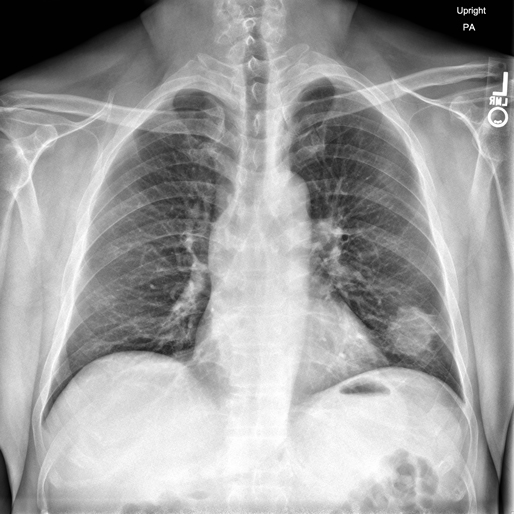
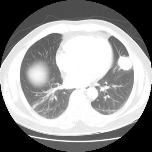
Plain films and a CT scan of the chest show a 3.5 cm rounded peripheral mass in the left upper lobe with possible left hilar lymphadenopathy. Further imaging reveals no other lesions in the abdomen, thorax, head, or neck. A CT-guided FNA of the lesion was performed along with a core needle biopsy.
Radiographs
Chest plain film and CT show a peripheral radiodense "coin-lesion" on the left.
Click on images to enlarge.
 |
 |
Microscopic Findings
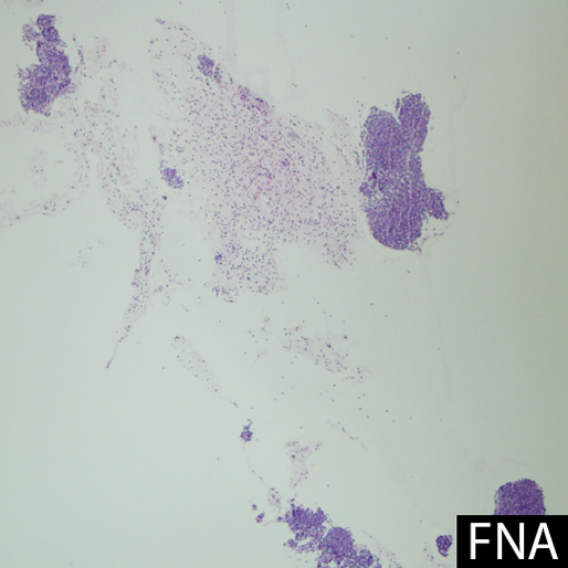
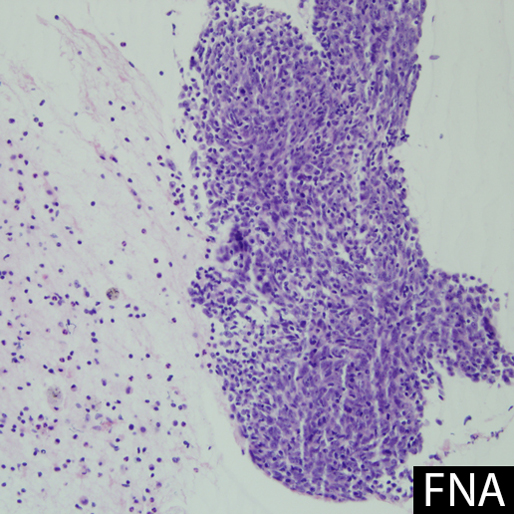
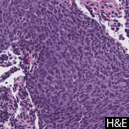
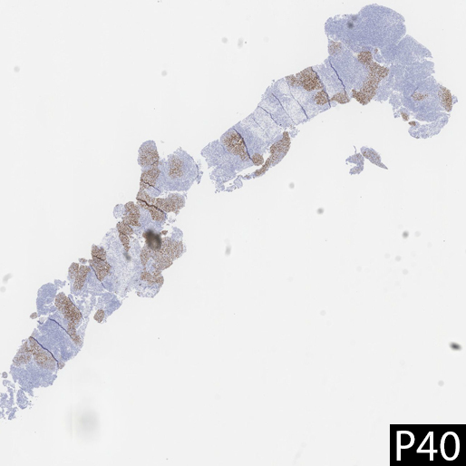
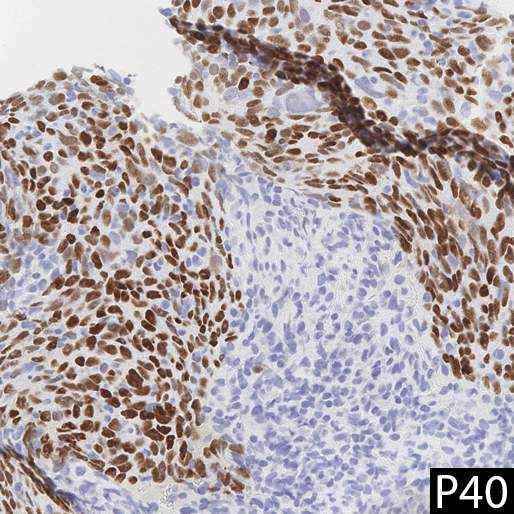
Sections of the core needle biopsy show sheets of large atypical epithelioid cells with prominent nucleoli, abundant cytoplasm, indistinct cell borders, and abundant surrounding chronic inflammatory cells (predominantly lymphocytes). By immunohistochemistry, the cells of interest are positive for AE1/AE3 and p40 (additional stains, including CD45, CK7, CK20, TTF-1, CD56, synaptophysin, and chromogranin, are negative). The aspirate smears show similar histologic findings.
FNA showed a cellular aspirate with cohesive groups of epitheloid cells intermixed with small mononuclear cells.
Click on images to enlarge.
 |
 |
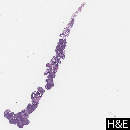
On H&E, the core needle biopsy shows a population of epithelioid cells with infiltrating small mononuclear cells, with some areas of peripheral lymphocytic aggregates.
Click on images to enlarge.
 |
 |
 |
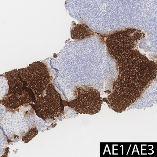
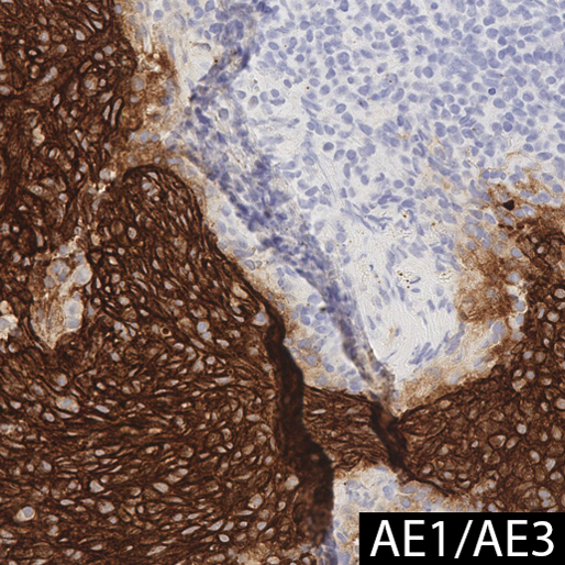
AE1/AE3 and P40 staining highlight the epithelioid cells, but not the background or infiltrating lymphocytes.
Click on images to enlarge.
 |
 |
 |
 |
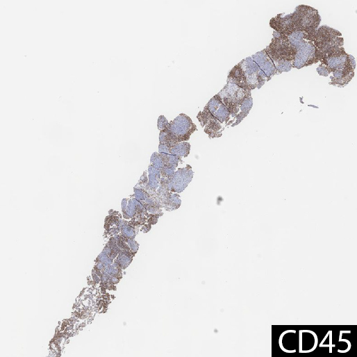
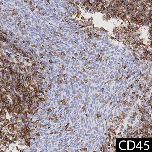
CD45 highlights the lymphocytes surrounding and infiltrating the epithelioid cells.
Click images to enlarge.
 |
 |
What is the diagnosis?
Choose one answer and submit.
 Meet our Residency Program Director