
Residency Program - Case of the Month
December 2016 - Presented by Nima Amini
Clinical History
A 75-year-old man presented with a 2 year history of a lesion on the left temple. A shave excision was performed on a 1.1 cm well-demarcated, ulcerated, brown pigmented plaque.
Microscopic Description
Microscopically, sections showed a nodular dermal proliferation of atypical spindle cells without infiltrative growth or subcutaneous involvement (Figure 1). The tumor cells were
- Negative: S100, Melan A, AE1/AE3, Desmin and CD34.
- Positive: CD10 (strong, Figure 3) and smooth muscle actin (SMA) (focal).
More than two years later, he presented with a left neck mass. A fine needle aspiration biopsy was performed on the secondary lesion. It showed an identical morphology and immunophenotype as the skin tumor (in addition, 34BE12, MNF116 and p63 were all negative), consistent with metastasis. (Figures 4,5,6).
Click to enlarge image.
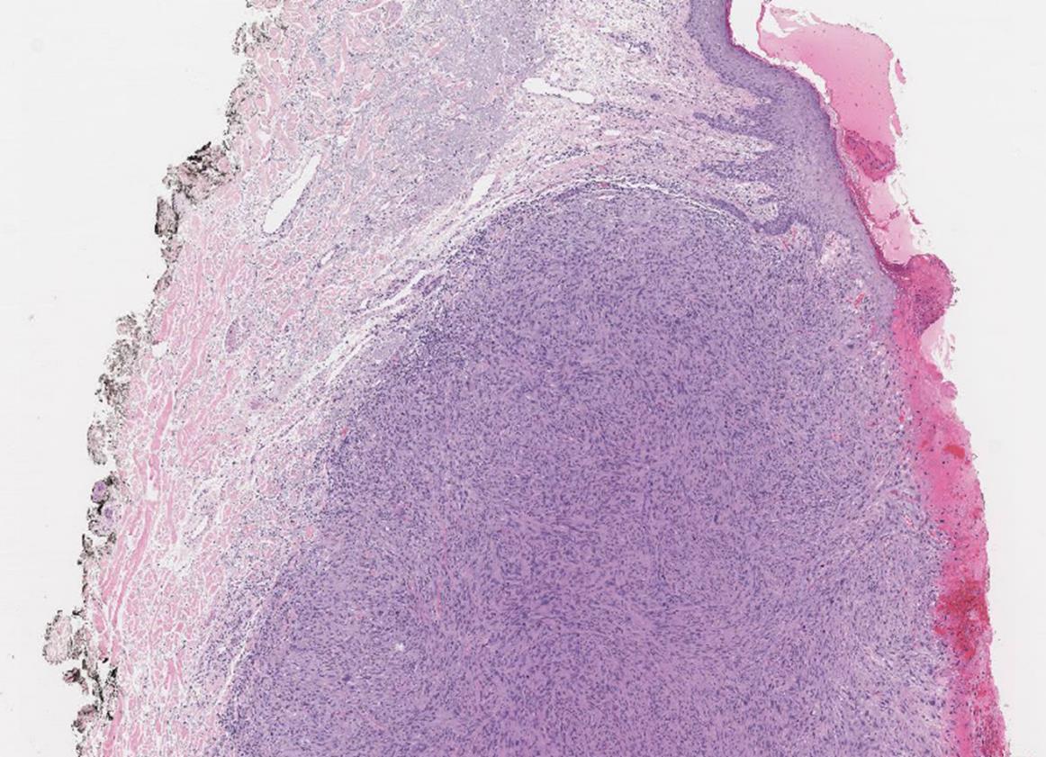
Figure 1
The well-demarcated, ulcerated primary lesion on H&E stain (20x)

Figure 2
Bizarre multinucleated tumor cells in hypercellular stroma with frequent mitotic figures, on H&E stain (200x)
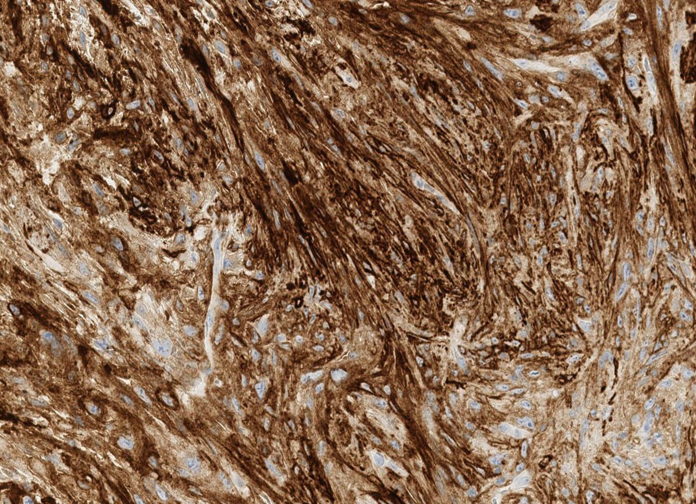
Figure 3
Primary lesion, tumor cells are strongly positive for CD10 (200x)
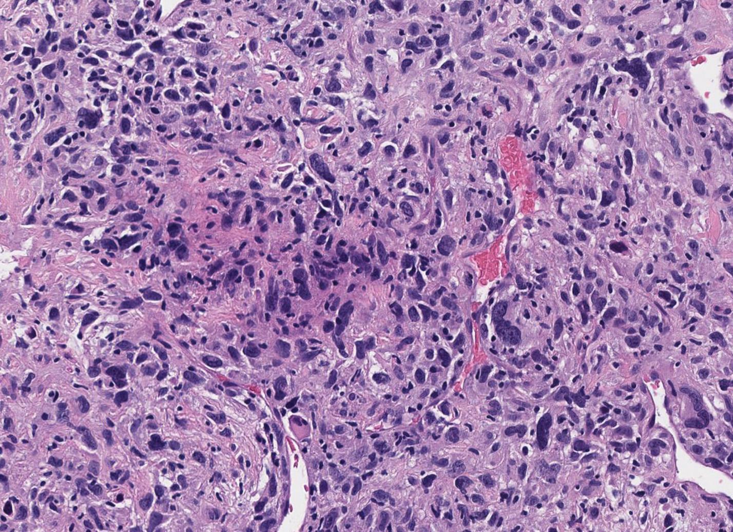
Figure 4
Identical histologic features seen in the secondary lesion on H&E stain (200x)

Figure 5
Secondary lesion, tumor cells are strongly positive for CD10 (200x)
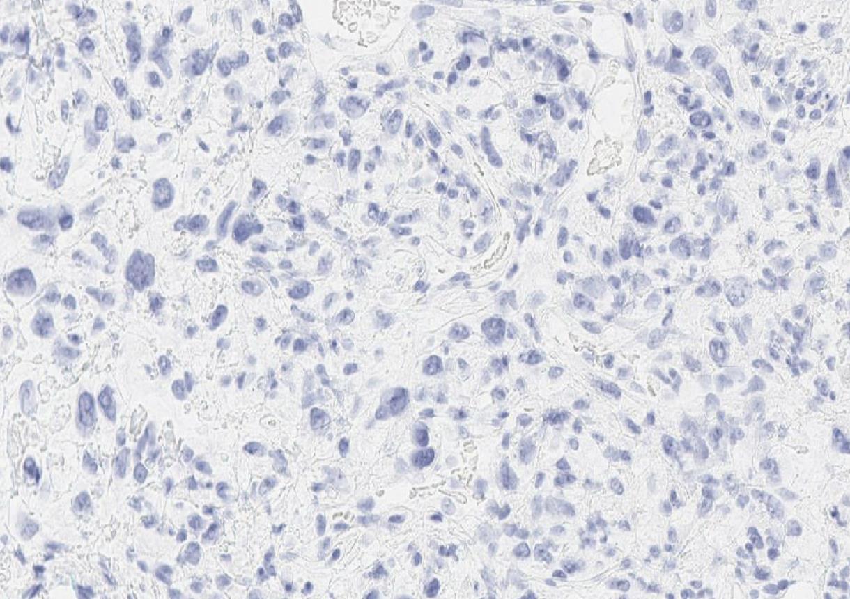
Figure 6
Tumor cells in secondary lesion are negative for MNF116 (200x)
What is the diagnosis?
Choose one answer and submit.
 Meet our Residency Program Director