
Resident Program - Case of the Month
February 2019 - Presented by Ryan Thomas (Mentored by Anthony Karnezis)
Discussion
Fetus papyraceus describes the phenomenon that occurs when a fetus in a multiple gestation pregnancy dies in utero and becomes compressed and mummified in the placental membranes. On gross examination, the compressed and flattened mass bears a resemblance to parchment paper, giving this unique entity its name. Fetal demise is thought to usually occur between 12 and 20 weeks’ gestation, which allows for the compression and mummification to occur. If the fetal demise occurs earlier in the pregnancy, the fetus is likely to be entirely resorbed, while fetal demise later in the pregnancy results in maceration1. The incidence of fetus papyraceus is approximately 1 in 200 twin pregnancies and 1 in 12,000 pregnancies overall2.
Fetus papyraceus has been reported in both monochorionic and dichorionic twin pregnancies. There are no associations with age or parity, but there appears to be an increased frequency with monochorionic pregnancies and with velamentous umbilical cord insertion2.
In most cases, fetus papyraceus does not lead to any harm to the surviving twin2,3. However, there are reports of associations with aplasia cutis congenita1, intestinal atresia, gastroschisis, and cardiac and pulmonary anomalies2,3,4.
The case presented here is complicated by the fact that the tissue in the demised twin had a 69 XXX karyotype and the placental disc showed hydropic villi, which is consistent with the diagnosis of partial hydatidiform mole5. It appears that the fetal tissue produced by this molar pregnancy underwent the same process that a demised twin with a normal karyotype would undergo to form the ovoid plaque seen grossly. Remarkably, the microscopic sections show almost perfectly symmetrical representations of the cartilaginous structures (Figure 4).
Click on image to enlarge.
Figure 4
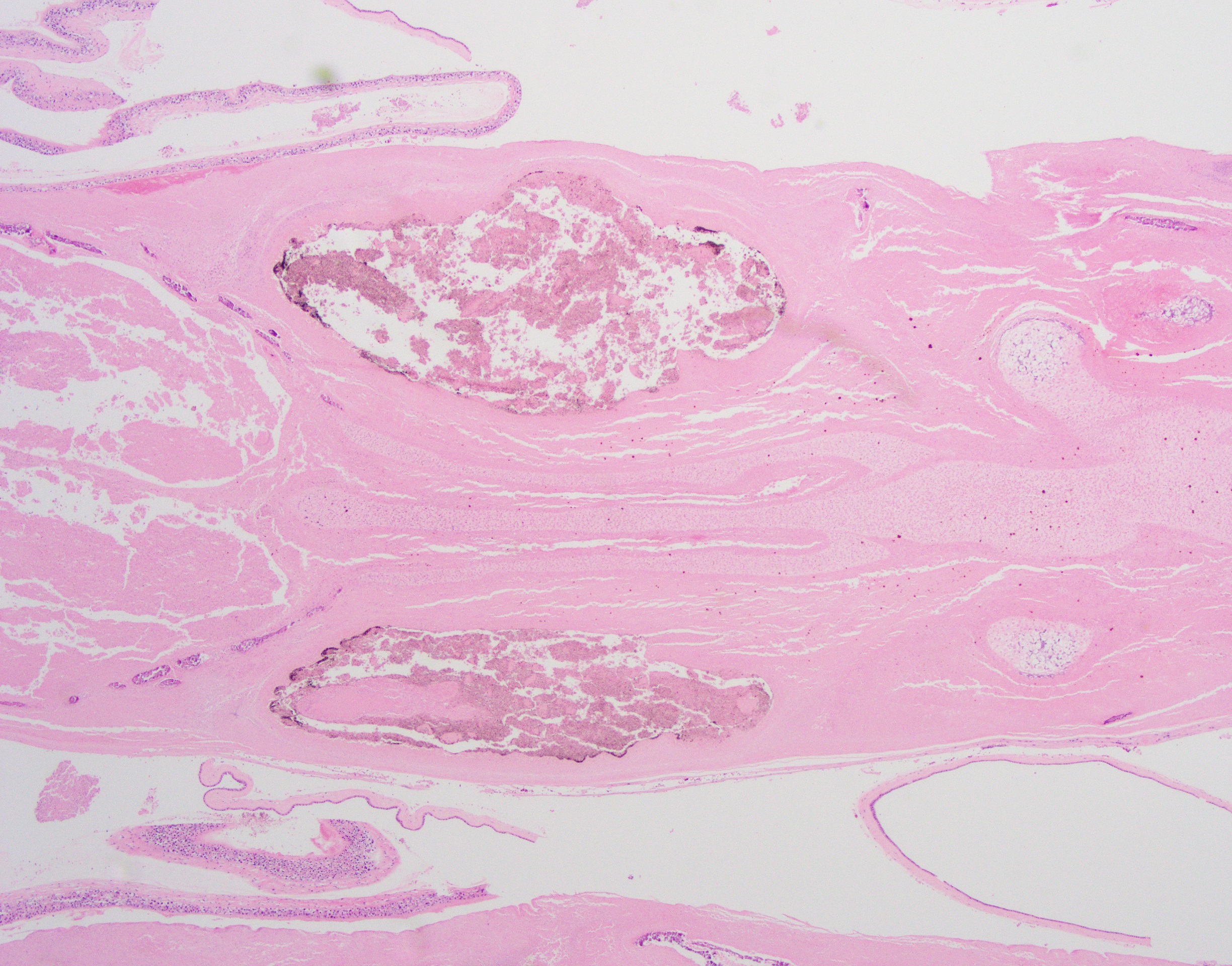
This image shows prominent symmetry of soft tissue structures and cartilaginous tissue. The structures were thought to represent brain with partially mineralized calvarium (left), eyes with melanin pigment (center), and immature cartilage of the bones of the face. (20X magnification).
References
- Pieretti, M., Alcalá, R., Boggio, P., Noguera-Morel, L., Porriño, M., & Luna, P. et al. (2015). "Aplasia Cutis Congenita Associated with Fetus Papyraceus". Pediatric Dermatology, 32(6), 858-861. doi: 10.1111/pde.12651
- McNamara, H., Kane, S., Craig, J., Short, R., & Umstad, M. (2016). "A review of the mechanisms and evidence for typical and atypical twinning". American Journal Of Obstetrics And Gynecology, 214(2), 172-191. doi: 10.1016/j.ajog.2015.10.930
- Dahiya, P., & Bains, R. (2014). "Conservative Management of Fetus Papyraceus: A Report of Two Cases". Oman Medical Journal, 29(2), 132-134. doi: 10.5001/omj.2014.32
- Matovelo, D., & Ndaboine, E. (2015). "Fetus papyraceus causing dystocia in a rural setting: a case report". Journal Of Medical Case Reports, 9(1). doi: 10.1186/s13256-015-0666-9
- Candelier, J. (2015). "The hydatidiform mole". Cell Adhesion & Migration, 10(1-2), 226-235. doi: 10.1080/19336918.2015.1093275
 Meet our Residency Program Director