Resident Program - Case of the Month
April 2019 - Presented by Raymond Gong (Mentored by Dorina Gui)
Clinical History
The patient is an 80-year old woman who presented with sudden onset of sharp, constant pain in her epigastrium that radiates to her chest. An abdominal ultrasound performed a few months prior was notable for multiple calculi, diffuse wall thickening, and a 0.5 cm polyp in the gallbladder.
After having reviewed her treatment options for symptomatic cholelithiasis, the patient elected for a laparoscopic cholecystectomy. Intra-operatively, a soft mass measuring approximately 1 cm was noted at the tip of the gallbladder fundus.
Gross Description
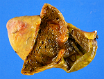
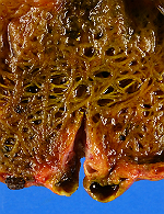
A soft nodule measuring 1.3 x 1.3 x 1.1 cm was located at the most distal tip of the gallbladder fundus. The gallbladder was opened to reveal a wall thickness measuring up to 0.5 cm, copious amounts of bile, and numerous calculi. The proximal third of the gallbladder showed unremarkable, velvety mucosa. The distal two-thirds of the gallbladder displayed a trabeculated wall and cystic cut surfaces. At the transition between trabeculated wall and normal mucosa was a circumferential stricture measuring 0.5 cm in thickness that reduced the luminal circumference to 0.6 cm (Figure 1). The fundic tip nodule was sectioned to reveal cystic cut surfaces filled with bile (Figure 2).
Click on image to enlarge.
Microscopic Description
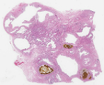
Sections of the trabeculated gallbladder wall demonstrated widespread herniations of the mucosa through discontinuous muscle bundles of the muscularis, which are commonly referred to as Rokitansky-Aschoff sinuses. The numerous intramural glands present in the specimen were often cystically dilated and filled with inspissated bile (Figures 3 and 4). Sections of the stricture and fundic nodule similarly showed the presence of dilated glands in the muscularis with extension into the subserosa (Figures 5 and 6). Significant architectural complexity was not identified. The epithelium lining the intramural glands varied from a simple cuboidal to columnar, pancreatobiliary type-epithelium to a metaplastic, pyloric type-epithelium consisting of columnar cells with abundant mucin vacuoles (Figure 7). In both types of epithelium, the nuclei were fairly uniform and polarized toward the basal periphery of the cells. There was no significant cytologic atypia such as pseudostratification, nuclear enlargement, nuclear irregularity, increased nucleus-to-cytoplasm ratio, mitoses, or loss of polarity.
Click on image to enlarge.
What is the diagnosis?
Choose one answer and submit.


 Meet our Residency Program Director
