
Resident Program - Case of the Month
May 2019 - Presented by Alison Chan (Mentored by Chihong Zhou)
Clinical History
The patient is a 32-year-old woman with a nine-month history of constitutional symptoms of ‘just not feeling good.’ She describes symptoms of abdominal malaise, epigastric discomfort that abruptly began without any triggers. She denies any vomiting, diarrhea, or melena. Her past medical history is significant for obesity. She does not drink, smoke, or use any illicit substances.
She was previously worked-up with their results as the following:
- Pregnancy test – negative
- Gastroesophageal reflux disease – given PPI
- H. pylori gastritis – negative
- Celiac disease – negative – was put on elimination and gluten-free diet
- CBC and CMP unremarkable
- Stool cultures – negative
Physical exam findings include normal bowel sounds, soft abdomen, no masses, or organomegaly. She is mildly sensitive over the epigastrium and left lower quadrant, but no guarding or rebound. No CVA tenderness.
Imaging
Ultrasonography, CT scan of the abdomen, and MRI imaging of the abdomen show a 7.8 x 5.7 x 3.5 cm well-defined, ovoid, retroperitoneal mass in the right upper quadrant. The lesion contacts the pancreas, right kidney, right adrenal gland, duodenum, and inferior vena cava, but does not invade into these organs. There is no connection to the biliary system. No other lesions are identified by imaging. A transduodenal ultrasound-guided fine needle aspiration of the lesion was performed.
CT and MRI
Click on images to enlarge.
Figure 1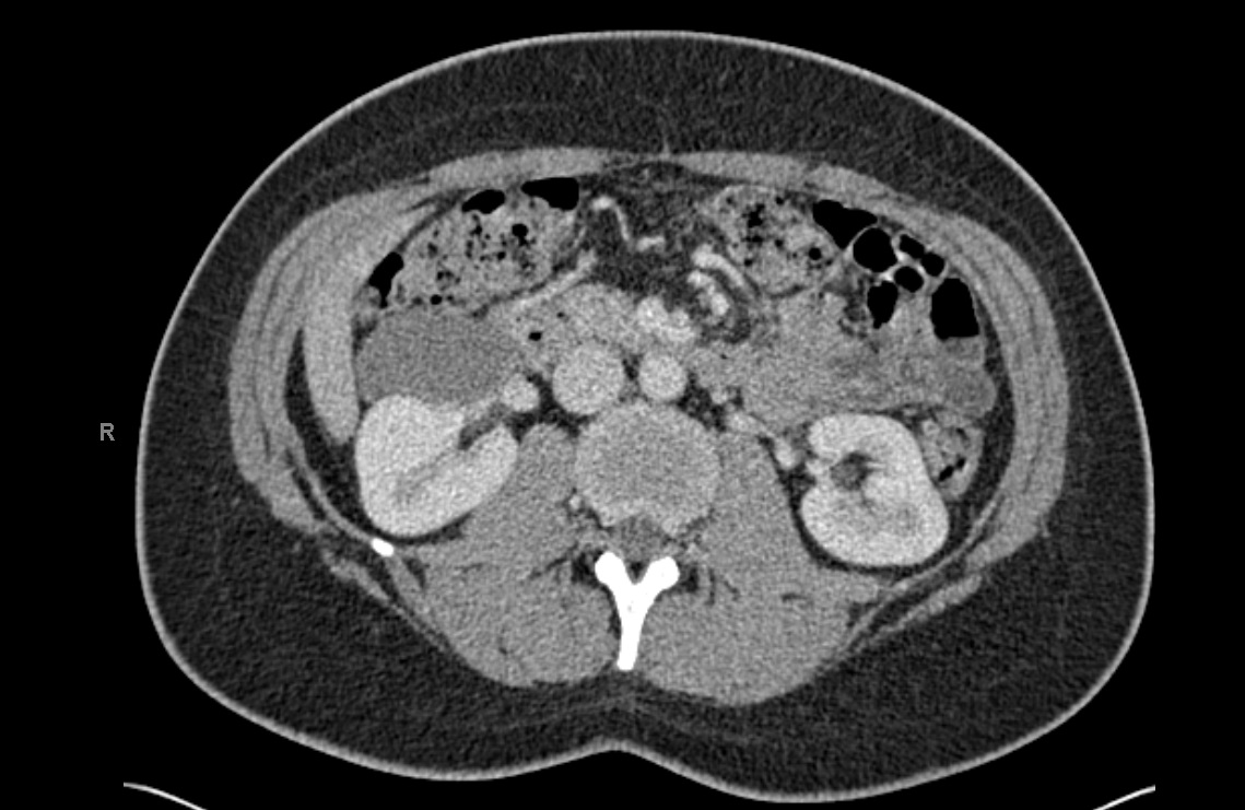
Figure 2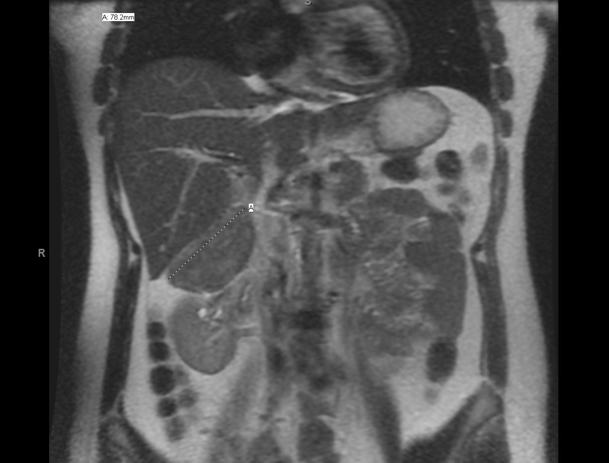
Figure 3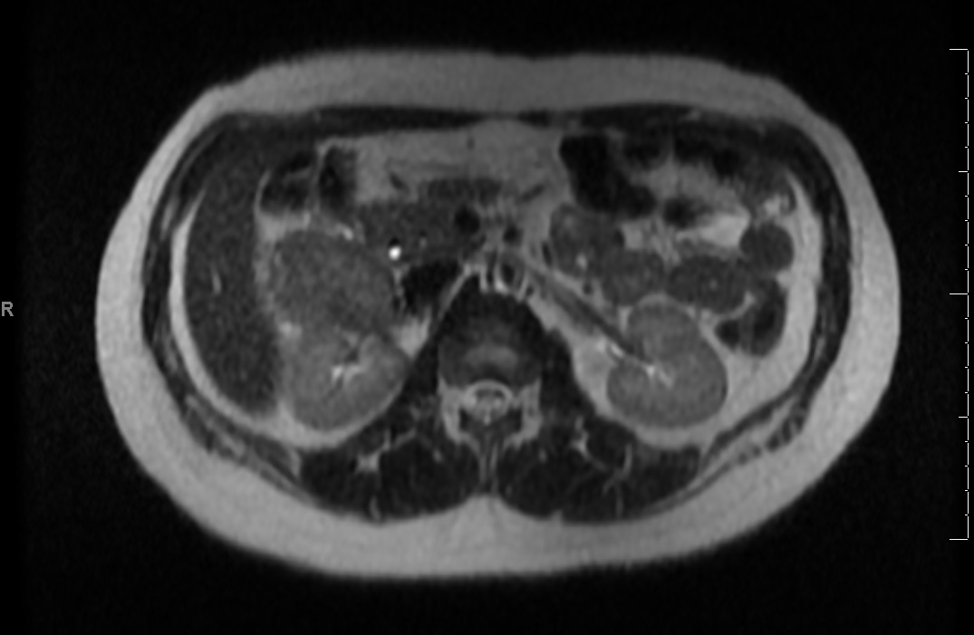
Microscopic Findings
The FNA smears are sparsely cellular consisting of sheets and groups of columnar cells, rare small fragments of spindle cells, mucus, and blood. The cell block is sparsely cellular consisting of rare fragments of spindled cells within a loose stroma, abundant sheets and groups of columnar cells, mucus, and blood. Immunohistochemistry was performed on the cell block.
Click on images to enlarge.
Figure 4: PAP (10x)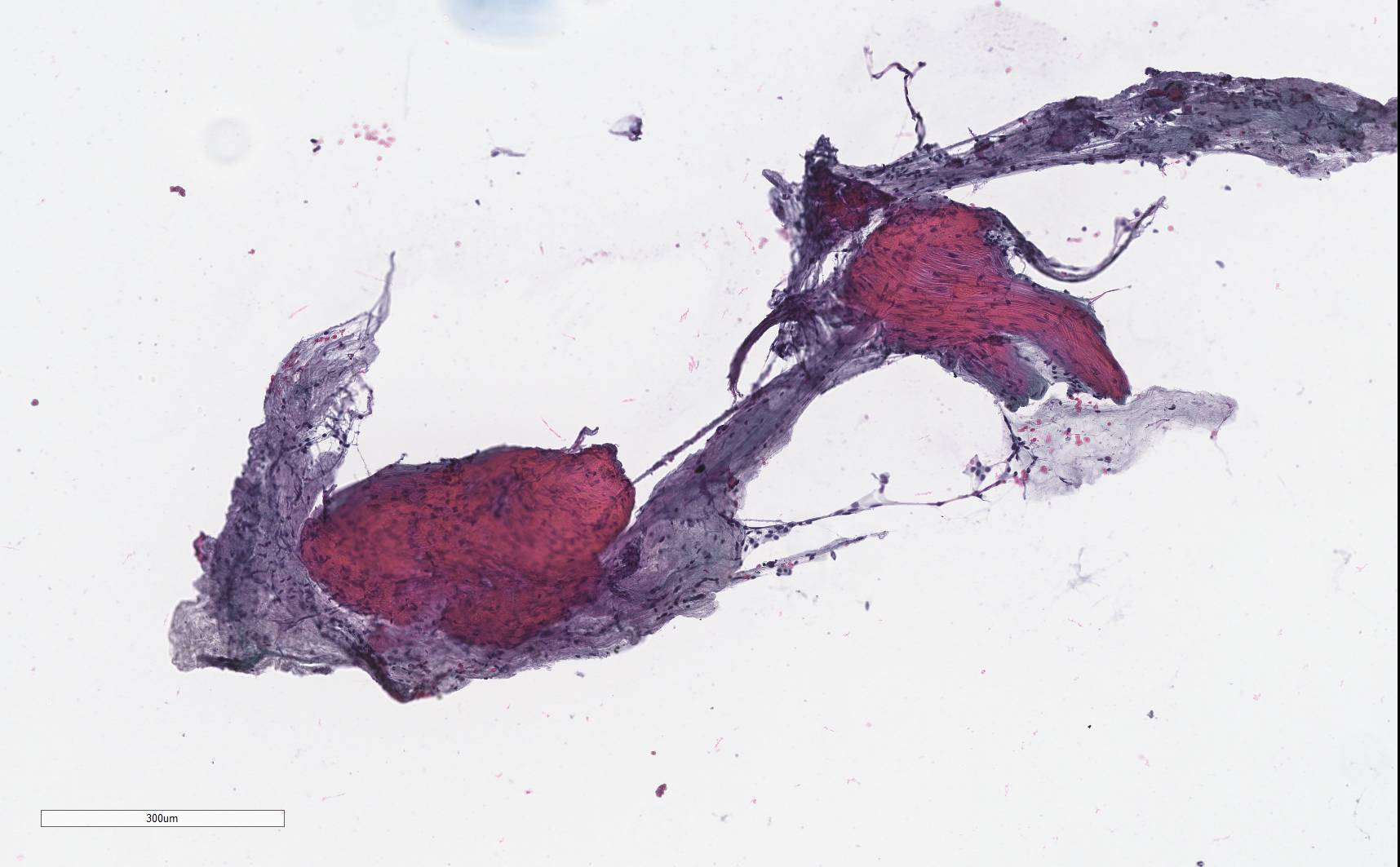
Figure 5: Giemsa (10x)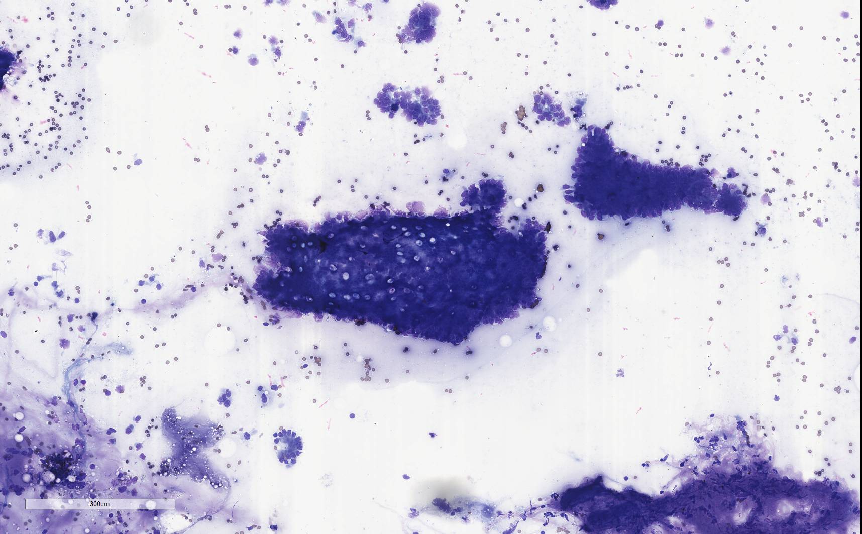
Figure 6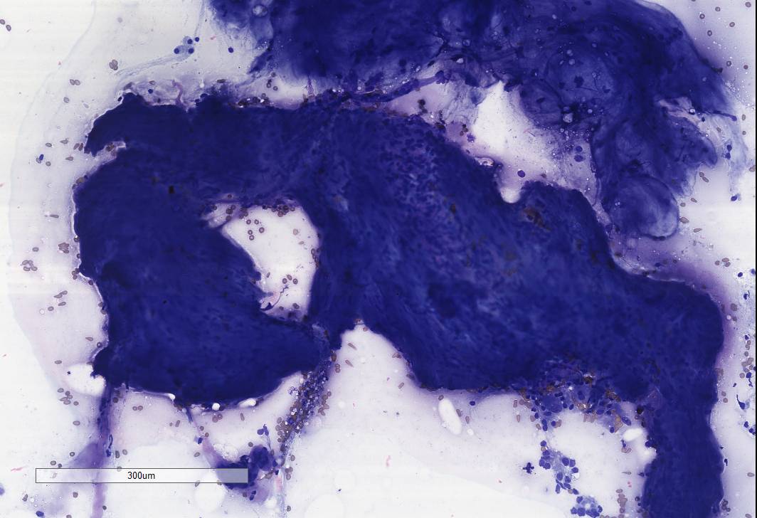
Figure 7: Cell Block 10x
Figure 8: Cell Block 20x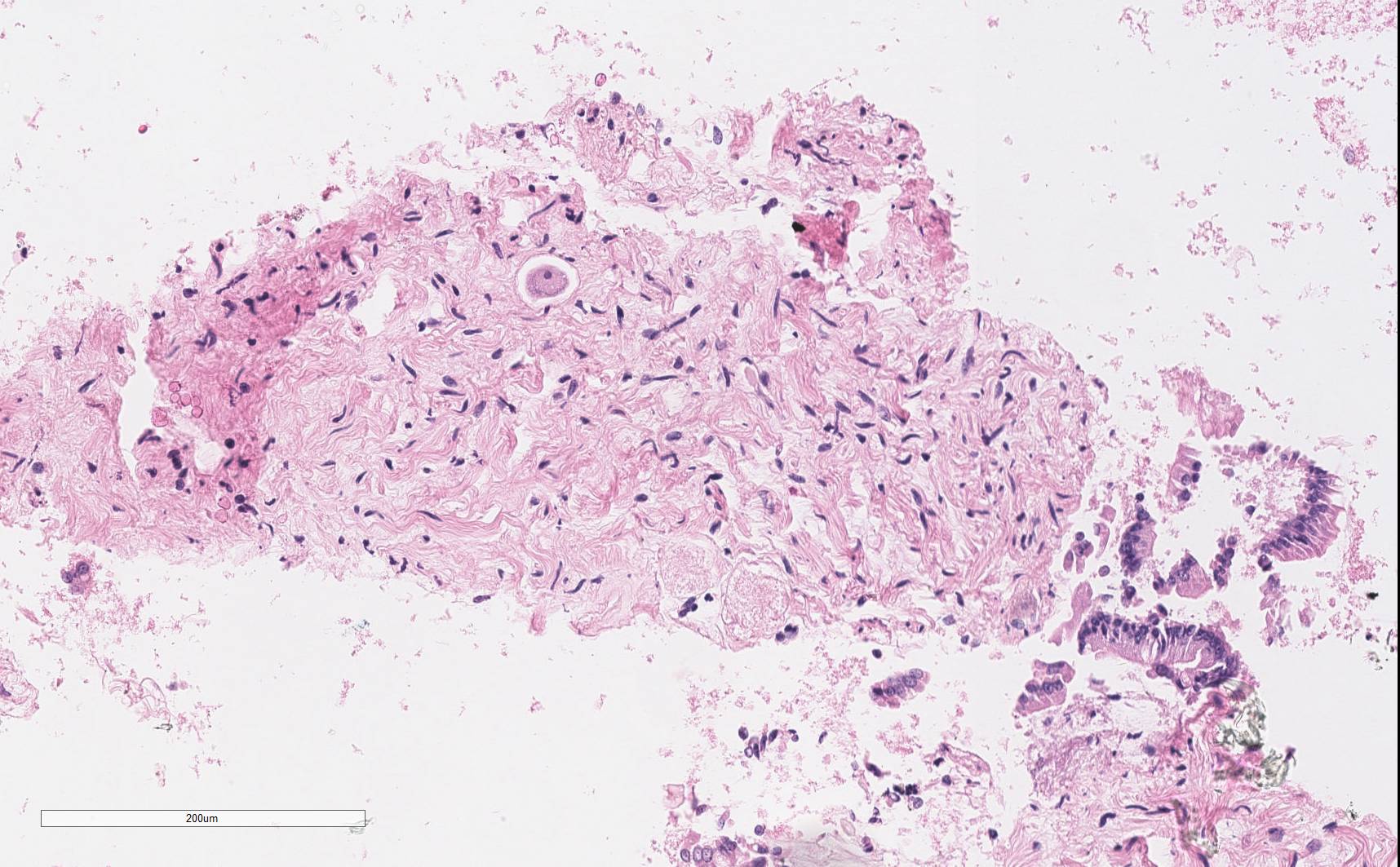
Figure 9: Immunohistochemical stains (all at 10)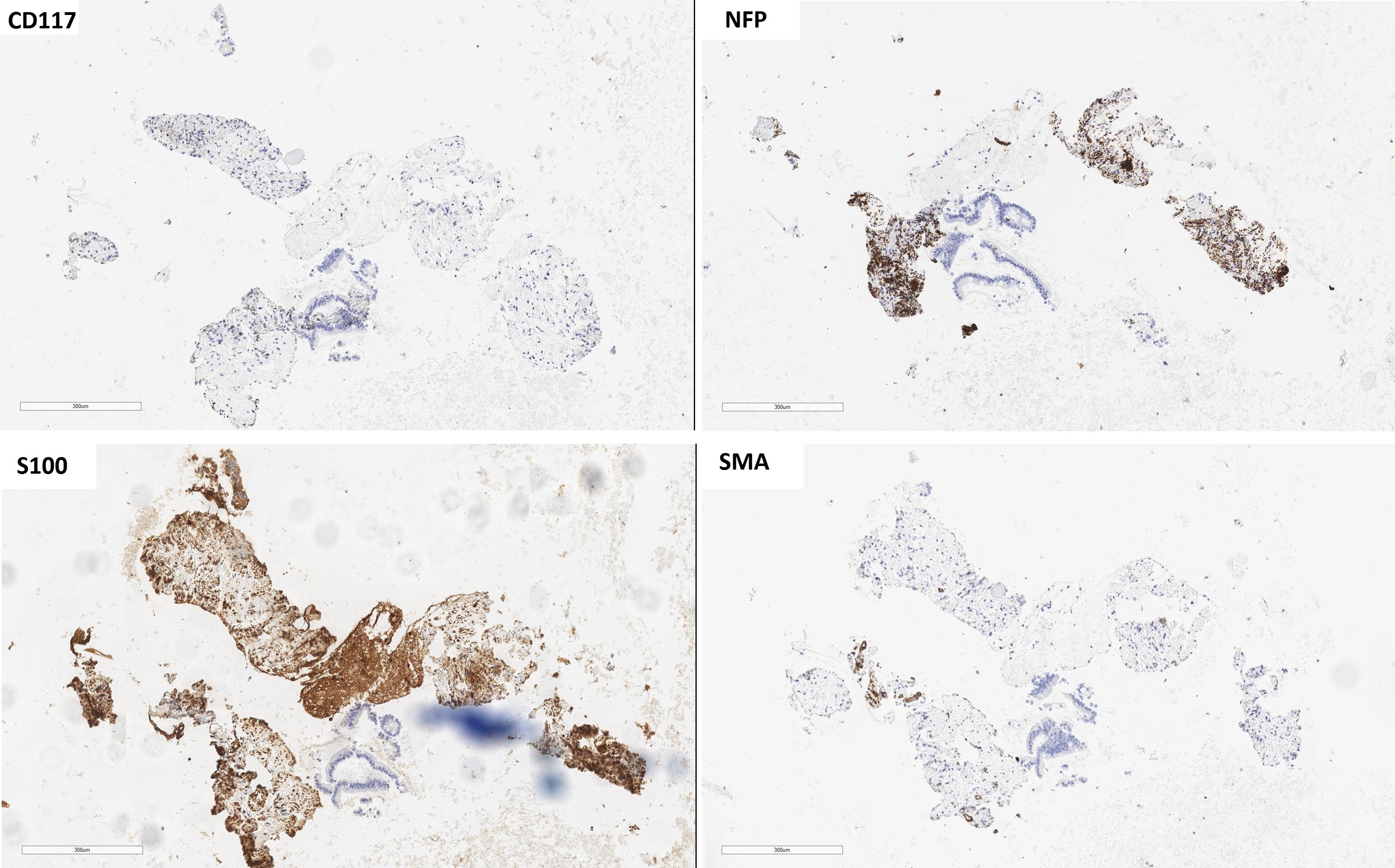
What is the diagnosis?
Choose one answer and submit.
 Meet our Residency Program Director