
Resident Program - Case of the Month
December 2019 - Presented by Alison Chan (Mentored by Regina Gandour-Edwards)
Clinical History:
The patient is an 85-year-old man with intermittent gross hematuria for the past few months. He reports intermittent symptoms of hesitancy and frequency. Four months ago, he underwent a transurethral resection of bladder tumor (TURBT) that identified two small tumors of unclear pathology. There was no subsequent therapy or cystoscopy done at that time. Currently, he denies any blood clots in his urine. Past medical history includes Chronic Kidney Disease 4 (CKD4), congestive heart failure, and chronic anemia. He is a former smoker with no known family history of genitourinary malignancies.
Physical exam findings showed mild abdominal distention, but otherwise unremarkable.
Imaging and Procedures:
One month ago, a CT scan showed a 2.5 cm lobular, vascular mass arising from the right posterior and superior wall of the urinary bladder that was suspicious for a neoplasm. There was additional marked diffuse irregular thickening of the bladder.
At this time, the TURBT showed large, multifocal bladder tumors that appeared invasive. The trigone was not involved. The clinician was unable to completely resect the tumor due to invasive disease and multiple bladder diverticuli. Tissue was obtained in this procedure for pathologic diagnosis.
Microscopic Findings:
The TURBT specimen consists of multiple fragments of tissue with papillary architecture and small to large nests of cells in an organoid fashion, admixed with large amounts of necrosis and hemorrhage. The cells are somewhat small and monotonous with scattered pleomorphic cells that vary in sizes containing hyperchromatic, irregularly shaped nuclei. The cells have a high nucleus to cytoplasmic ratio with nuclear molding. The nuclear chromatin is predominantly in a finely dispersed with occasional prominent nucleoli. A few immunohistochemical stains are performed, with the results depicted below in the slide images.
Click on images to enlarge.

Figure 1: H&E at 2x magnification.
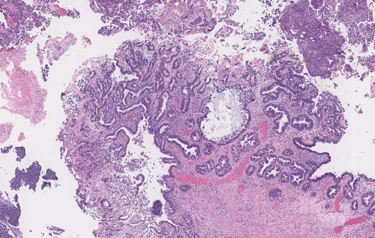
Figure 2: H&E at 4x magnification.
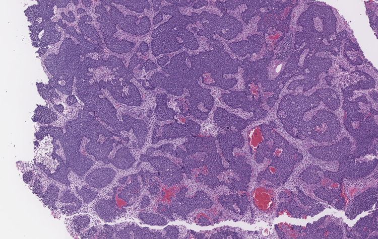
Figure 3: H&E at 4x magnification.
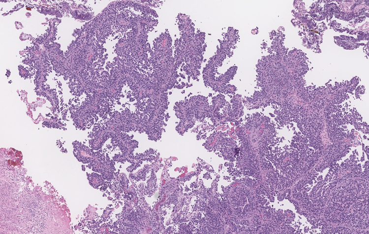
Figure 4: H&E at 4x magnification.
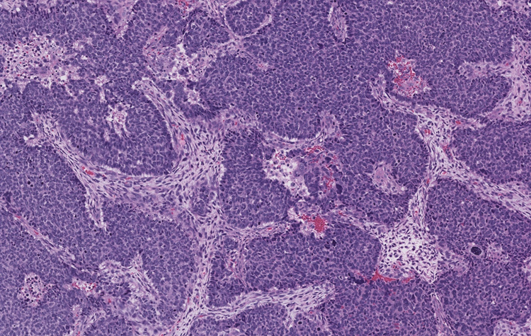
Figure 5: H&E at 10x magnification.
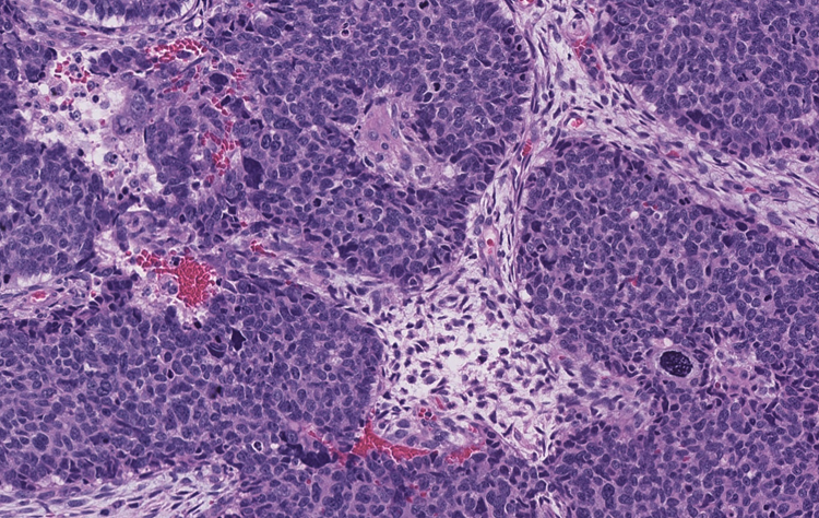
Figure 6: H&E at 20x magnification.
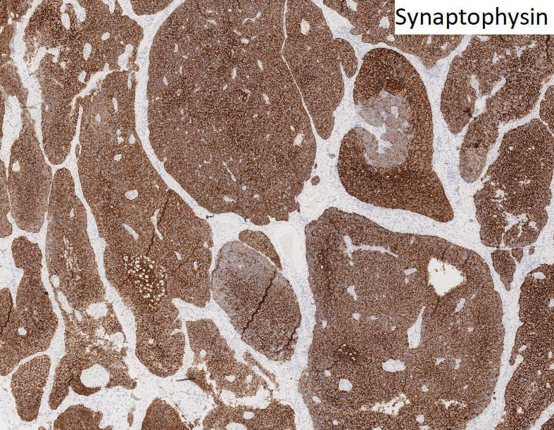
Figure 7: Immunohistochemical stains.
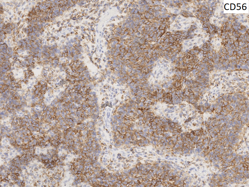
Figure 8: Immunohistochemical stains.
 Meet our Residency Program Director