
November 2021 – Presented by Dr. Alexander Ladenheim (Mentored by Dr. Han Lee)
Clinical History
A male in his 60s with a history of chronic rhinosinusitis and prior functional endoscopic sinus surgeries presents with a 3.8 cm expansile lesion eroding into the clivus.
Microscopic Findings
The tumor cells are arranged in sheets and nest-like areas without associated brain parenchyma. The cells are monotonous and contain scant, pink cytoplasm with ill-defined cell borders. Their nuclei are hyperchromatic with fine chromatin, indistinct nucleoli, and occasional mitoses. Frequent nuclear pseudoinclusions are present.
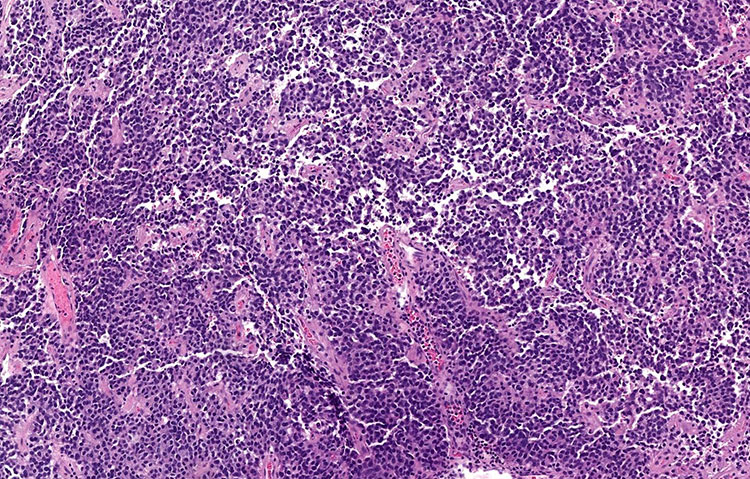
Figure 1: Sheets and nests of monotonous, polygonal cells (H&E, 100x).
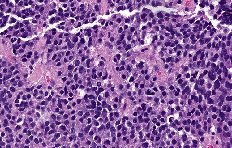
Figure 2: The cells contain round nuclei with smudgy, finely granular chromatin; a nuclear pseudoincluion is seen in this field (H&E, 400x).
 Meet our Residency Program Director