
March 2022 – Presented by Dr. Jiejun Wu (Mentored by Dr. Han S. Lee)
The patient is a 72 year old male with progressively worsening low back pain and lower extremity symptoms consistent with neurogenic claudication. Contrast lumbar spine MRI showed a 7 mm intradural extramedullary homogenously enhancing lesion at L1/L2 level within the posterior thecal sac. He denies history of cancer. He is treated with laminectomy and neoplasm resection. The received specimen designated as “intradural L1 tumor” is a 0.9 x 0.8 x 0.3 cm soft tissue fragment which is bisected and entirely submitted as follows (Fig. 1-2).
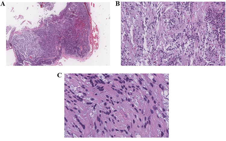
Figure 1. Intradural L1 tumor. H&E. A, 20x. B, 100x. C, 200x
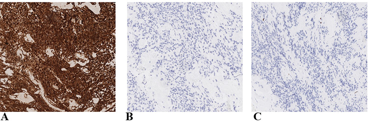
Figure 2. IHC, 100x. A, GFAP. B, EMA. C Ki67.
 Meet our Residency Program Director